File:Phytomyxea collage.jpg
Jump to navigation
Jump to search

Size of this preview: 501 × 600 pixels. Other resolutions: 200 × 240 pixels | 600 × 718 pixels.
Original file (600 × 718 pixels, file size: 201 KB, MIME type: image/jpeg)
Captions
Captions
Add a one-line explanation of what this file represents
Summary[edit]
| Description |
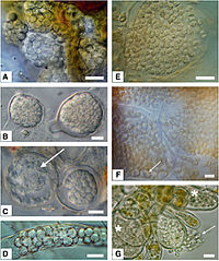
English: Morphology of resting spores from selected phytomyxids. Bar=10 μm.
|
| Date | |
| Source | Neuhauser S., Kirchmair M., Bulman S., Bass D. (2014). "Cross-kingdom host shifts of phytomyxid parasites". BMC Evolutionary Biology 14 (33). DOI:10.1186/1471-2148-14-33. |
| Author | Sigrid Neuhauser, Martin Kirchmair, Simon Bulman and David Bass |
Licensing[edit]
This file is licensed under the Creative Commons Attribution 2.0 Generic license.
|
This file was published in a BioMed Central journal. Their website states that all of its research publications is published under the license which is identical to the Creative Commons Attribution 2.0 license (some non-research articles like reviews or editorials may require a subscription.)
To the uploader: You must provide a link (URL) to the original file or journal article.
|
File history
Click on a date/time to view the file as it appeared at that time.
| Date/Time | Thumbnail | Dimensions | User | Comment | |
|---|---|---|---|---|---|
| current | 13:38, 24 January 2016 | | 600 × 718 (201 KB) | Mithril (talk | contribs) | =={{int:filedesc}}== {{Information |Description= {{en|1=Morphology of resting spores from selected phytomyxids. Bar=10 μm: '''left column''': Plasmodiophorida, '''right column''': Phagomyxida. *'''A''': ''Sorosphaerula viticola'': hollow sporosori in... |
You cannot overwrite this file.
File usage on Commons
The following page uses this file:
File usage on other wikis
The following other wikis use this file:
- Usage on ar.wikipedia.org
- Usage on arz.wikipedia.org
- Usage on en.wikipedia.org
- Usage on es.wikipedia.org
- Usage on ia.wikipedia.org
- Usage on ko.wikipedia.org
- Usage on pl.wikipedia.org
- Usage on ro.wikipedia.org
- Usage on ru.wikipedia.org
- Usage on tr.wikipedia.org
- Usage on www.wikidata.org