Category:Endoplasmic reticulum
Перейти к навигации
Перейти к поиску
органелла эукариотических клеток  | |||||
| Медиафайл | |||||
| Это частный случай понятия |
| ||||
|---|---|---|---|---|---|
| Подкласс от |
| ||||
| Является частью | |||||
| Соединяется с |
| ||||
| |||||
Подкатегории
В этой категории отображается 4 подкатегории из имеющихся 4.
Файлы в категории «Endoplasmic reticulum»
Показаны 200 файлов из 201, находящегося в данной категории.
(Предыдущая страница) (Следующая страница)-
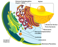
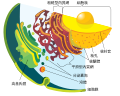
 0313 Endoplasmic Reticulum a en.png 604 × 362; 230 Кб
-
 0313 Endoplasmic Reticulum b en.png 467 × 363; 190 Кб
-
 0313 Endoplasmic Reticulum b labeled.png 371 × 363; 196 Кб
-
 0313 Endoplasmic Reticulum c en.png 733 × 398; 245 Кб
-
 0313 Endoplasmic Reticulum c labeled.png 604 × 398; 250 Кб
-
 0313 Endoplasmic Reticulum.jpg 1102 × 892; 488 Кб
-
 201601 Endoplasmic reticulum.png 400 × 400; 92 Кб
-
 Blausen 0350 EndoplasmicReticulum ar.png 1440 × 1054; 1,21 Мб
-
 Blausen 0350 EndoplasmicReticulum.png 1600 × 1054; 1,24 Мб
-
 Cellorganeller pic swe 27-05-2007.png 603 × 481; 100 Кб
-
 Clara cell lung - TEM.jpg 640 × 480; 98 Кб
-
 Drawing of Endoplasmic Reticulum.jpg 2435 × 1807; 1,19 Мб
-
 Endomembrane system diagram cs.svg 638 × 530; 326 Кб
-
 Endomembrane system diagram de.svg 612 × 486; 96 Кб
-
 Endomembrane system diagram en.svg 612 × 486; 109 Кб
-
 Endomembrane system diagram es.svg 662 × 506; 95 Кб
-
 Endomembrane system diagram fr.svg 612 × 486; 244 Кб
-
 Endomembrane system diagram hu.svg 638 × 530; 326 Кб
-
 Endomembrane system diagram id.svg 640 × 500; 188 Кб
-
 Endomembrane system diagram it.svg 612 × 486; 79 Кб
-
 Endomembrane system diagram ka.svg 612 × 486; 89 Кб
-
 Endomembrane system diagram ku .svg 612 × 486; 88 Кб
-
 Endomembrane system diagram ln.svg 640 × 500; 188 Кб
-
 Endomembrane system diagram nl.svg 765 × 600; 192 Кб
-
 Endomembrane system diagram notext.svg 560 × 460; 98 Кб
-
 Endomembrane system diagram pl.svg 612 × 486; 249 Кб
-
 Endomembrane system diagram pt.svg 612 × 486; 286 Кб
-
 Endomembrane system diagram ru.svg 612 × 486; 78 Кб
-
 Endomembrane system diagram tr.svg 612 × 486; 163 Кб
-
 Endomembrane system diagram uk.svg 614 × 498; 295 Кб
-
 Endomembrane system diagram zh-tw.svg 612 × 486; 80 Кб
-
 Endomembrane system diagram zh.svg 612 × 486; 87 Кб
-
 Endoplasmic reticulum - ribosomes.jpg 1221 × 788; 356 Кб
-
 Endoplasmic reticulum -- Smart-Servier.jpg 10 240 × 5760; 2,51 Мб
-
 Endoplasmic reticulum 1 -- Smart-Servier.png 1755 × 835; 284 Кб
-
 Endoplasmic reticulum 2 -- Smart-Servier.png 1898 × 723; 329 Кб
-
 Endoplasmic reticulum 3 -- Smart-Servier.png 1900 × 666; 282 Кб
-
 Endoplasmic reticulum 4 -- Smart-Servier.png 1760 × 843; 334 Кб
-
 Endoplasmic reticulum 5 -- Smart-Servier.png 1749 × 459; 126 Кб
-
 Endoplasmic reticulum 6 -- Smart-Servier.png 2117 × 674; 223 Кб
-
 Endoplasmic reticulum 7 -- Smart-Servier.png 2118 × 611; 170 Кб
-
 Endoplasmic reticulum 8 -- Smart-Servier.png 1753 × 490; 161 Кб
-
-
-
ER-network-dynamics-are-differentially-controlled-by-myosins-XI-K-XI-C-XI-E-XI-I-XI-1-and-XI-2-Movie1.ogv 4,3 с, 1280 × 720; 2,11 Мб
-
-
ER-network-dynamics-are-differentially-controlled-by-myosins-XI-K-XI-C-XI-E-XI-I-XI-1-and-XI-2-Movie3.ogv 4,3 с, 1280 × 720; 1,88 Мб
-
ER-network-dynamics-are-differentially-controlled-by-myosins-XI-K-XI-C-XI-E-XI-I-XI-1-and-XI-2-Movie4.ogv 4,5 с, 1280 × 720; 2,25 Мб
-
ER-network-dynamics-are-differentially-controlled-by-myosins-XI-K-XI-C-XI-E-XI-I-XI-1-and-XI-2-Movie5.ogv 4,3 с, 1280 × 720; 1,33 Мб
-
ER-network-dynamics-are-differentially-controlled-by-myosins-XI-K-XI-C-XI-E-XI-I-XI-1-and-XI-2-Movie6.ogv 4,3 с, 1280 × 720; 1,38 Мб
-
ER-network-dynamics-are-differentially-controlled-by-myosins-XI-K-XI-C-XI-E-XI-I-XI-1-and-XI-2-Movie7.ogv 4,3 с, 1280 × 720; 1,45 Мб
-
ER-network-dynamics-are-differentially-controlled-by-myosins-XI-K-XI-C-XI-E-XI-I-XI-1-and-XI-2-Movie8.ogv 4,3 с, 1280 × 720; 1,25 Мб
-
-
 Esquema de la teoria de l'endosimbio siseriada.png 4408 × 6476; 2,96 Мб
-
Exogenous-Ether-Lipids-Predominantly-Target-Mitochondria-pone.0031342.s006.ogv 28 с, 480 × 360; 8,69 Мб
-
-
-
-
-
-
-
-
-
-
-
-
-
-
-
-
-
-
Induction-of-protein-body-formation-in-plant-leaves-by-elastin-like-polypeptide-fusions-1741-7007-7-48-S4.ogv 6,0 с, 1106 × 1099; 2,77 Мб
-
-
-
Induction-of-protein-body-formation-in-plant-leaves-by-elastin-like-polypeptide-fusions-1741-7007-7-48-S7.ogv 5,0 с, 1106 × 1099; 3,55 Мб
-
Induction-of-protein-body-formation-in-plant-leaves-by-elastin-like-polypeptide-fusions-1741-7007-7-48-S8.ogv 5,0 с, 1106 × 1099; 1,36 Мб
-
-
Inhibition-of-the-Unfolded-Protein-Response-Mechanism-Prevents-Cardiac-Fibrosis-pone.0159682.s006.ogv 1 м 14 с, 640 × 368; 39,48 Мб
-
Inside-out-Ca2+-signalling-prompted-by-STIM1-conformational-switch-ncomms8826-s2.ogv 5,3 с, 1160 × 448; 559 Кб
-
Inside-out-Ca2+-signalling-prompted-by-STIM1-conformational-switch-ncomms8826-s3.ogv 5,0 с, 752 × 564; 208 Кб
-
Inside-out-Ca2+-signalling-prompted-by-STIM1-conformational-switch-ncomms8826-s4.ogv 5,0 с, 1004 × 504; 288 Кб
-
Inside-out-Ca2+-signalling-prompted-by-STIM1-conformational-switch-ncomms8826-s5.ogv 6,0 с, 1176 × 592; 89 Кб
-
-
-
-
-
-
-

-
-
Mitochondrial-pleomorphy-in-plant-cells-is-driven-by-contiguous-ER-dynamics-Video1.ogv 20 с, 290 × 184; 1,39 Мб
-
Mitochondrial-pleomorphy-in-plant-cells-is-driven-by-contiguous-ER-dynamics-Video2.ogv 3,8 с, 548 × 192; 618 Кб
-
Mitochondrial-pleomorphy-in-plant-cells-is-driven-by-contiguous-ER-dynamics-Video3.ogv 7,3 с, 368 × 258; 970 Кб
-
Mitochondrial-pleomorphy-in-plant-cells-is-driven-by-contiguous-ER-dynamics-Video4.ogv 21 с, 392 × 602; 1,98 Мб
-
Mitochondrial-pleomorphy-in-plant-cells-is-driven-by-contiguous-ER-dynamics-Video5.ogv 48 с, 386 × 246; 4,3 Мб
-
Mitochondrial-pleomorphy-in-plant-cells-is-driven-by-contiguous-ER-dynamics-Video6.ogv 45 с, 378 × 146; 3,25 Мб
-
Molecular-Determinants-and-Dynamics-of-Hepatitis-C-Virus-Secretion-ppat.1002466.s016.ogv 6,2 с, 270 × 235; 145 Кб
-
Molecular-Determinants-and-Dynamics-of-Hepatitis-C-Virus-Secretion-ppat.1002466.s017.ogv 10 с, 476 × 347; 584 Кб
-
Molecular-Determinants-and-Dynamics-of-Hepatitis-C-Virus-Secretion-ppat.1002466.s018.ogv 3,1 с, 291 × 230; 141 Кб
-
Molecular-Determinants-and-Dynamics-of-Hepatitis-C-Virus-Secretion-ppat.1002466.s019.ogv 6,7 с, 216 × 234; 391 Кб
-
Molecular-Determinants-and-Dynamics-of-Hepatitis-C-Virus-Secretion-ppat.1002466.s020.ogv 5,0 с, 82 × 118; 83 Кб
-
Molecular-Determinants-and-Dynamics-of-Hepatitis-C-Virus-Secretion-ppat.1002466.s021.ogv 9,6 с, 240 × 283; 166 Кб
-
-
Molecular-Determinants-and-Dynamics-of-Hepatitis-C-Virus-Secretion-ppat.1002466.s023.ogv 4,1 с, 65 × 89; 26 Кб
-
Molecular-Determinants-and-Dynamics-of-Hepatitis-C-Virus-Secretion-ppat.1002466.s024.ogv 9,3 с, 58 × 58; 42 Кб
-
-
Molecular-Determinants-and-Dynamics-of-Hepatitis-C-Virus-Secretion-ppat.1002466.s026.ogv 4,0 с, 68 × 74; 21 Кб
-
Molecular-Determinants-and-Dynamics-of-Hepatitis-C-Virus-Secretion-ppat.1002466.s027.ogv 2,5 с, 11 × 52; 10 Кб
-
-
-
Molecular-Determinants-and-Dynamics-of-Hepatitis-C-Virus-Secretion-ppat.1002466.s030.ogv 5,4 с, 38 × 50; 19 Кб
-
Motion-and-Flexibility-in-Human-Cytochrome-P450-Aromatase-pone.0032565.s011.ogv 56 с, 480 × 360; 3,85 Мб
-
Motion-and-Flexibility-in-Human-Cytochrome-P450-Aromatase-pone.0032565.s012.ogv 1 м 5 с, 480 × 360; 4,04 Мб
-
Motion-and-Flexibility-in-Human-Cytochrome-P450-Aromatase-pone.0032565.s013.ogv 52 с, 480 × 360; 6,07 Мб
-
Motion-and-Flexibility-in-Human-Cytochrome-P450-Aromatase-pone.0032565.s014.ogv 1 м 5 с, 640 × 360; 15,66 Мб
-
Motion-and-Flexibility-in-Human-Cytochrome-P450-Aromatase-pone.0032565.s015.ogv 1 м 36 с, 480 × 360; 8,03 Мб
-
Motion-and-Flexibility-in-Human-Cytochrome-P450-Aromatase-pone.0032565.s016.ogv 1 м 38 с, 480 × 360; 6,03 Мб
-
Motion-and-Flexibility-in-Human-Cytochrome-P450-Aromatase-pone.0032565.s017.ogv 2 м 51 с, 480 × 360; 14,38 Мб
-
 N-linked protein glycosylation in the ER.svg 2662 × 1018; 164 Кб
-
Nuclear-envelope-associated-endosomes-deliver-surface-proteins-to-the-nucleus-ncomms9218-s2.ogv 26 с, 768 × 576; 4,38 Мб
-
Nuclear-envelope-associated-endosomes-deliver-surface-proteins-to-the-nucleus-ncomms9218-s3.ogv 36 с, 768 × 576; 2,56 Мб
-
Nuclear-envelope-associated-endosomes-deliver-surface-proteins-to-the-nucleus-ncomms9218-s4.ogv 19 с, 640 × 480; 6,58 Мб
-
Nuclear-envelope-associated-endosomes-deliver-surface-proteins-to-the-nucleus-ncomms9218-s5.ogv 13 с, 768 × 576; 4,99 Мб
-
 Nucleus ER golgi ex.jpg 534 × 426; 49 Кб
-
 Nucleus ER golgi.svg 492 × 565; 168 Кб
-
 Nucleus ER.png 416 × 469; 31 Кб
-
 Pancreatic acinar cells - TEM.jpg 640 × 480; 160 Кб
-
 Pathologies liées aux modifications du réticulum sarcoplasmique.jpg 704 × 449; 120 Кб
-
 Phosphatidylinositol hydrolysis and synthesis.jpg 6000 × 4200; 2,65 Мб
-
 PLC.png 621 × 706; 276 Кб
-
 ProteinTranscription+Synthesis.svg 957 × 531; 82 Кб
-
-
-
-
-
-
-
-
-
-
-
-
-
-
-
-
 RER Gland MO.png 1019 × 766; 1,02 Мб
-
 Reticle-endoplasmatic.jpg 499 × 291; 53 Кб
-
 Reticulo 3D Coanofl.png 786 × 408; 360 Кб
-
 Reticulo dinámica Levadu.png 1340 × 1748; 1,03 Мб
-
 Reticulo Endoplasmico. Inmunodetección RFP-RPGR..png 856 × 257; 359 Кб
-
 Reticulo Levadura.png 357 × 432; 189 Кб
-
 Reticulo Nanodominio.png 1200 × 343; 273 Кб
-
 Reticulo periferico.png 514 × 754; 378 Кб
-
 Reticulo sectores Levad.png 835 × 1341; 951 Кб
-
 Retikulum stanica.png 500 × 540; 97 Кб
-
 Retículo central Levadura.png 358 × 435; 229 Кб
-
 RETÍCULO ENDOPLASMÁTICO.jpg 180 × 239; 37 Кб
-
 Retîkûlûma Endoplazmî.png 762 × 418; 221 Кб
-
 Rough endoplasmic reticulum.JPG 471 × 278; 33 Кб
-
 Rough ER Close up.png 1188 × 672; 138 Кб
-
-
-
-
-
-
 Smooth Endoplasmic Reticulum.jpg 1195 × 549; 194 Кб
-
Spatial-Temporal-Study-of-Rab1b-Dynamics-and-Function-at-the-ER-Golgi-Interface-pone.0160838.s002.ogv 37 с, 1920 × 1080; 7,13 Мб
-
-
Spatial-Temporal-Study-of-Rab1b-Dynamics-and-Function-at-the-ER-Golgi-Interface-pone.0160838.s004.ogv 50 с, 1280 × 720; 4,59 Мб
-
-
-
-
-
-
-
-
-
-
-
-
-
-
-
-
-
-
-
The-Human-Polyoma-JC-Virus-Agnoprotein-Acts-as-a-Viroporin-ppat.1000801.s008.ogv 9,1 с, 180 × 110; 99 Кб
-
-
-
-
-
The-Intracellular-Transport-and-Secretion-of-Calumenin-12-in-Living-Cells-pone.0035344.s005.ogv 5,1 с, 512 × 512; 5,14 Мб
-
The-Intracellular-Transport-and-Secretion-of-Calumenin-12-in-Living-Cells-pone.0035344.s006.ogv 5,1 с, 512 × 512; 10,09 Мб
-
The-Intracellular-Transport-and-Secretion-of-Calumenin-12-in-Living-Cells-pone.0035344.s007.ogv 5,0 с, 512 × 512; 6,93 Мб
-
The-Intracellular-Transport-and-Secretion-of-Calumenin-12-in-Living-Cells-pone.0035344.s008.ogv 5,0 с, 512 × 512; 13,77 Мб
-
The-Intracellular-Transport-and-Secretion-of-Calumenin-12-in-Living-Cells-pone.0035344.s009.ogv 5,0 с, 512 × 512; 5,2 Мб
-
The-Intracellular-Transport-and-Secretion-of-Calumenin-12-in-Living-Cells-pone.0035344.s010.ogv 5,0 с, 512 × 512; 5,49 Мб


