File:Echinococcus vogeli2.jpg
(Redirigé depuis File:Ev2.jpg)
Taille de cet aperçu : 800 × 526 pixels. Autres résolutions : 320 × 210 pixels | 640 × 421 pixels | 1 024 × 673 pixels | 1 280 × 841 pixels | 3 059 × 2 010 pixels.
Fichier d’origine (3 059 × 2 010 pixels, taille du fichier : 1,65 Mio, type MIME : image/jpeg)
Légendes
Légendes
Ajoutez en une ligne la description de ce que représente ce fichier
| Description |
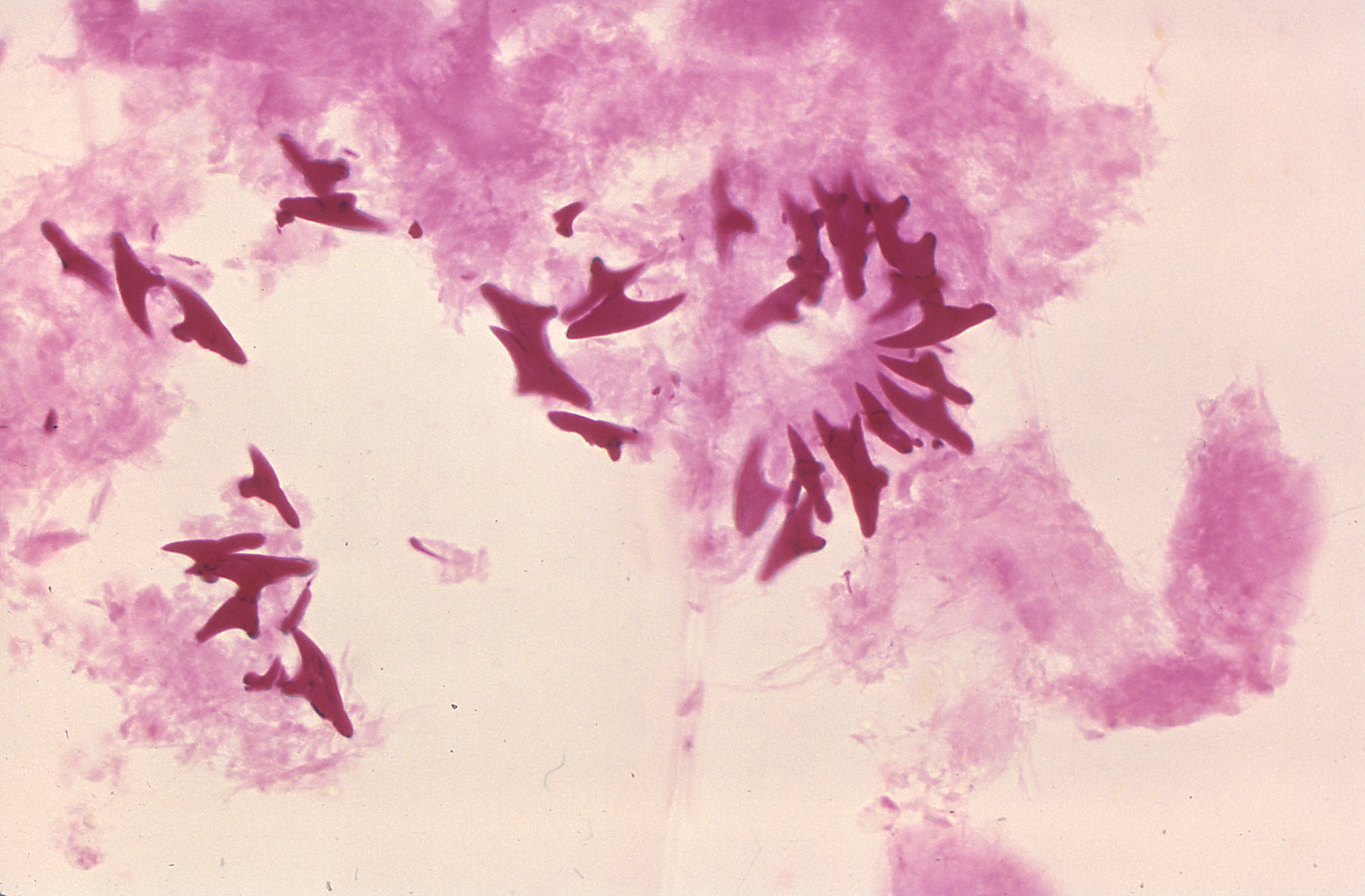
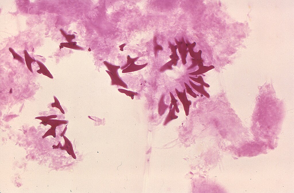
English: This photomicrograph of a tissue sample harvested from a cyst, which was harbored inside a gorilla, revealed the presence of these protoscolex hooks of a microscopic Echinococcus vogeli tapeworm. The larval stage of this microscopic tapeworm is one of the causative agents of alveolar hydatid disease (AHD), an infection in humans that causes parasitic tumors to form, mainly in the liver, but can also appear in other organs as well. |
||
| Date | |||
| Source |
|
||
| Auteur | CDC/Dr. Peter Schantz |
Cette image est l’œuvre des
Centers for Disease Control and Prevention , division du Département de la Santé et des Services Sociaux des États-Unis, réalisée par un employé dans le cadre de ses activités professionnelles. En tant qu'œuvre du gouvernement fédéral des États-Unis d'Amérique, cette image est placée dans le domaine public.
|
Historique du fichier
Cliquer sur une date et heure pour voir le fichier tel qu'il était à ce moment-là.
| Date et heure | Vignette | Dimensions | Utilisateur | Commentaire | |
|---|---|---|---|---|---|
| actuel | 4 août 2020 à 07:08 | | 3 059 × 2 010 (1,65 Mio) | TommyG (d | contributions) | Larger version from source |
| 4 août 2020 à 05:19 |  | 700 × 477 (46 kio) | WQUlrich (d | contributions) | Reverted to version as of 13:34, 18 November 2007 (UTC) | |
| 12 mars 2008 à 15:56 |  | 417 × 488 (178 kio) | EraserGirl (d | contributions) | rmv border | |
| 27 février 2008 à 08:31 |  | 640 × 480 (63 kio) | Musaraigne (d | contributions) | {{Information |Description=Eugène Viala dans son atelier |Source=travail personnel |Date=2007 |Author= Musaraigne |Permission= |other_versions= }} | |
| 18 novembre 2007 à 13:34 |  | 700 × 477 (46 kio) | Filip em (d | contributions) | {{Information |Description=This is a photomicrograph of protoscolex hooks of Echinococcus vogeli taken from a cyst within a gorilla. The larval stage of the microscopic tapeworm Echinococcus vogeli is one of the causative agents of Alveolar Hydatid Disea |
Vous ne pouvez pas remplacer ce fichier.
Utilisations locales du fichier
La page suivante utilise ce fichier :
- File:Ev2.jpg (redirection de fichier)