Category:Active site
Jump to navigation
Jump to search
active site of an enzyme; region of an enzyme where substrate molecules bind and undergo a chemical reaction to form products  | |||||
| Upload media | |||||
| Instance of | |||||
|---|---|---|---|---|---|
| Part of | |||||
| |||||
English: Region of an enzyme where substrate molecules bind and undergo a chemical reaction.
Media in category "Active site"
The following 148 files are in this category, out of 148 total.
-
 1-3 BPG in active site.png 341 × 415; 38 KB
-
 1FIW Active Site Residues.png 4,524 × 2,859; 2.25 MB
-
 1FIW Active Site Surface.png 4,524 × 2,859; 3.11 MB
-
 4cysresi.png 829 × 514; 269 KB
-
 4PGI-A.png 1,106 × 430; 362 KB
-
 Acetoacetate decarboxylase biounit 3BH3 with inset.png 2,032 × 2,018; 2.41 MB
-
 Acetylene Hydratase from Pelobacter acetylenicus active site.png 1,044 × 765; 288 KB
-
 AChe active site.jpg 333 × 193; 9 KB
-
 ACS Active site.png 1,324 × 1,250; 785 KB
-
 Actie Site of Eflornithine.png 494 × 515; 223 KB
-
 Actieve centrum.jpg 1,600 × 984; 192 KB
-
 Active Site 1.png 491 × 366; 72 KB
-
 Active site ADH1.png 590 × 500; 110 KB
-
 Active site bacterial DIC.png 607 × 498; 297 KB
-
 Active Site close up of tryptophan 7 halogenase.png 1,696 × 1,288; 482 KB
-
 ACTIVE SITE Isopenicillin-N synthase.JPG 676 × 428; 46 KB
-
 Active site mutations.jpeg 354 × 528; 43 KB
-
 Active site NagA.png 960 × 575; 399 KB
-
 Active site of ASL with argininosuccinate.png 1,264 × 722; 520 KB
-
 Active site of CAII (cropped).jpg 1,472 × 1,090; 138 KB
-
 Active site of CAII.jpg 2,242 × 1,266; 227 KB
-
 Active Site of Cyanophycinase.png 1,280 × 980; 630 KB
-
 Active site of GAPDH of E. coli.png 2,400 × 2,400; 1.68 MB
-
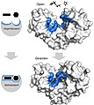
 Active Site of Gelonin with Adenine.jpg 876 × 691; 87 KB
-
 Active site of glucansucrase in Lactobacillus reuteri.png 640 × 434; 103 KB
-
 Active site of human ACE2.png 2,400 × 2,400; 806 KB
-
 Active site of mouse GPI.png 2,400 × 2,400; 835 KB
-
 Active site of Thermus thermophilus argininosuccinate synthetase 01.png 996 × 714; 257 KB
-
 Active site of urease.png 304 × 195; 7 KB
-
 Active Site with labeled amino acids and NAD.png 859 × 600; 286 KB
-
 Activesite PEPPM.jpg 400 × 400; 49 KB
-
 Activesite101.png 924 × 324; 242 KB
-
 ActiveSiteLaccase-2.png 2,791 × 1,625; 2.01 MB
-
 ActiveSitesCorrected.png 4,052 × 913; 230 KB
-
 ADAR1 active site.png 636 × 462; 55 KB
-
 Allosteric comp inhib 1.svg 840 × 580; 29 KB
-
 Allosteric comp inhib 2.svg 840 × 580; 27 KB
-
 Allosteric Regulation.svg 512 × 384; 11 KB
-
 AN20758251-28-082-g001.jpg 740 × 355; 70 KB
-
 ASL active sites.png 640 × 480; 445 KB
-
 ASPA bound to an intermediate analogue.png 588 × 514; 90 KB
-
 Aspartyl Protease Active Site.gif 527 × 569; 10 KB
-
 Binding of MgATP by nitrogenase.png 513 × 321; 12 KB
-
 CAMP.gif 300 × 299; 55 KB
-
 Carbonic anhydrase 1CA2 active site.png 1,240 × 1,240; 445 KB
-
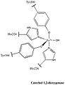 Catechol 1,2-dioxygenase.jpg 253 × 328; 17 KB
-
 CDO active site.png 3,057 × 1,523; 864 KB
-
 Centro activo aconitasa.jpg 1,146 × 643; 67 KB
-
 CENTRO ACTIVO.jpg 640 × 640; 89 KB
-
 Cholesterol-24 hydroxylase (CYP46A1) with active site in red.png 640 × 434; 82 KB
-
 Close-UpofLaccaseActiveSite.png 2,582 × 1,496; 1.93 MB
-
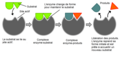
 ComplejoEnzimaSustrato.jpg 655 × 214; 54 KB
-
 Coordination in Active site.png 478 × 640; 54 KB
-
 CP Active Site.png 1,602 × 925; 1.48 MB
-

-
 Cyanophycinase active site image.png 1,458 × 1,126; 837 KB
-
 Cyanophycinase Active Site.png 1,450 × 1,124; 837 KB
-
 DAPDC Active Site.png 438 × 435; 49 KB
-
 Deoxyhemocyanin full.png 421 × 272; 9 KB
-
 DHFR methotrexate inhibitor.png 914 × 861; 176 KB
-
 DHFR methotrexate inhibitor.svg 1,800 × 1,728; 310 KB
-
 Diagramma adattamento indotto.svg 648 × 253; 18 KB
-

-
 DNA Pol II Active Site (PDB ID 3K5M).png 640 × 434; 111 KB
-
 DNA Pol II Active Site.png 640 × 434; 193 KB
-
 Docking.png 500 × 158; 9 KB
-
 Eflornithine in the Active Site.png 325 × 537; 321 KB
-
 Enolase active site.jpg 1,000 × 620; 55 KB
-
 Enzimműködés.jpg 800 × 346; 52 KB
-
 Enzyme action.png 1,247 × 634; 55 KB
-
 Enzyme structure (unannotated).svg 1,800 × 1,107; 465 KB
-
 Enzyme structure 2.svg 1,200 × 738; 815 KB
-
 Enzyme structure(rus).png 1,280 × 787; 325 KB
-
 Enzyme structure.png 1,361 × 837; 304 KB
-
 Enzyme structure.svg 1,800 × 1,107; 820 KB
-
 EnzymeActiveandAllostericSites.jpeg 650 × 316; 26 KB
-
 Fe-Ni hydrogenase activesite oxidizedandreduced.png 441 × 169; 14 KB
-
 Fech-2qd1-activesite.png 997 × 955; 166 KB
-
 Figure 3 modified.jpg 490 × 402; 38 KB
-
 Fpls-02-00028-g010.png 3,320 × 2,380; 2.14 MB
-
 GAD67 active site.png 910 × 641; 557 KB
-
 Gelonin Active Site.jpg 1,158 × 460; 74 KB
-
 Glucocerebrosidase active site.png 1,210 × 754; 578 KB
-
 Glucose-6-phosphate isomerase mechanism.svg 2,823 × 1,691; 134 KB
-
 Glycolytic Active site.png 300 × 260; 46 KB
-
 HDC Active Site Diagram.tif 960 × 720; 357 KB
-
 Hexokinase induced fit 2.svg 1,200 × 1,338; 1.3 MB
-
 Hexokinase induced fit.jpg 1,265 × 1,412; 253 KB
-
 Hexokinase induced fit.png 1,500 × 1,661; 737 KB
-
 Hexokinase induced fit.svg 1,500 × 1,673; 1.3 MB
-
 Histidine brace.tif 735 × 430; 23 KB
-
 Homoserine Dehydrogenase Active site.jpg 493 × 405; 30 KB
-
 Hsp90 ATP pocket.jpg 622 × 494; 42 KB
-
 Human APRTase, adenine binding site.png 661 × 485; 88 KB
-
 Induced fit diagram fr.png 2,008 × 980; 144 KB
-
 Induced fit diagram id.svg 648 × 253; 122 KB
-
 Induced fit diagram w. Swe. captions.png 648 × 253; 34 KB
-
 Induced fit diagram.png 648 × 253; 20 KB
-
 Induced fit diagram.svg 648 × 253; 21 KB
-
 Inducedfit080(rus).png 642 × 407; 37 KB
-
 Inducedfit080.png 642 × 407; 33 KB
-

-
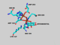
 Key residues that interact with ADP within the active site.png 574 × 714; 191 KB
-
 KinEnzym01.svg 490 × 270; 58 KB
-
 Labeled active site of 2aqj PrnA halogenase.png 2,088 × 1,374; 598 KB
-
 LaccaseActiveSite3.png 2,666 × 1,627; 2.01 MB
-
 Lock and key enzyme mechanism.svg 498 × 1,182; 18 KB
-
 Lock and key(rus).png 648 × 407; 35 KB
-
 Lock and key.png 648 × 407; 31 KB
-
 Malate dehydrogenase active site.svg 445 × 261; 41 KB
-
 Malate synthase structure including active site.png 1,650 × 1,275; 721 KB
-
 MDH-E-coli-AS.jpg 1,800 × 1,361; 211 KB
-
 Miox final 1.png 640 × 480; 55 KB
-
 Modèle de l'activité d'une enzyme.PNG 735 × 1,037; 79 KB
-
 Morphinone reductase active site structure.png 571 × 346; 33 KB
-
 MTHFR active site.jpg 939 × 649; 149 KB
-
 Nagalase-ActiveSite.png 2,400 × 2,400; 630 KB
-
 NDM-1 active site 4HL2.jpg 1,280 × 615; 120 KB
-
 Nitrilase Active Site.png 1,060 × 705; 376 KB
-
 NPPActiveSite.png 1,282 × 792; 109 KB
-
 Oxyhemocyanin full.png 316 × 239; 9 KB
-
 OxyHemoglobin active site (PDB code 2DN1).png 326 × 499; 41 KB
-
 PETase active site.png 891 × 416; 567 KB
-
 Pfprol active site.png 451 × 299; 63 KB
-
 Picture of Active Site 1Y79.png 1,590 × 943; 608 KB
-
 PMMO active site.png 2,400 × 1,627; 262 KB
-
 PNMT Active Site.png 1,599 × 900; 1.07 MB
-
 Porcine Mitochondrial IDH active site waters.jpg 1,162 × 859; 42 KB
-
 Possible Omega-amidase Active Site.png 640 × 434; 110 KB
-
 Proposed LAP-A active site.png 515 × 380; 18 KB
-
 Proximal Residues Bound To Citrate in Active Site of PGM.png 3,200 × 2,400; 2.27 MB
-
 Pymolactivesite.jpg 585 × 400; 48 KB
-
 Quin active site copy.jpg 520 × 388; 65 KB
-
 Quinacrine mustard in Trypanothione reductase active site.png 800 × 582; 292 KB
-
 RebH PyrH overlay.png 2,400 × 2,400; 591 KB
-
 Reverse transcriptase 3KLF labels-es.png 1,600 × 1,397; 1.17 MB
-
 Reverse transcriptase 3KLF labels.png 1,600 × 1,397; 1.52 MB
-
 RNaseH active site.svg 1,091 × 786; 118 KB
-
 RPE65 Active Site.png 980 × 566; 205 KB
-
 RuBisCOActiveSite.png 1,280 × 864; 305 KB
-
 SENP1 sitio catalítico.png 1,366 × 768; 668 KB
-
 SENP1.png 1,222 × 767; 789 KB
-
 Serum Paraoxonase-1 Active Site With Residue Labels.png 661 × 439; 182 KB
-
 TD PLP Site 2.png 1,098 × 897; 407 KB
-
 Topoisomerase IB Active Site.jpg 872 × 842; 59 KB
-
 Tryptophan-7-Halogenase Active Site.png 1,600 × 1,093; 837 KB
-
 Unión de PTX-2 a la Actina.jpg 514 × 246; 47 KB
-
 Usp27x active site.jpg 992 × 646; 110 KB


.jpg)


_with_active_site_in_red.png)


.png)


.svg)

.png)


.png)


.png)


.png)


