Category:Adenosine triphosphate
Jump to navigation
Jump to search
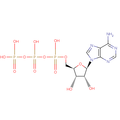
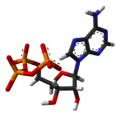
the energy-carrying molecule in living cells | |||||
| Upload media | |||||
| Instance of |
| ||||
|---|---|---|---|---|---|
| Subclass of |
| ||||
| Part of |
| ||||
| Has use | |||||
| Has part(s) | |||||
| Time of discovery or invention |
| ||||
| Physically interacts with |
| ||||
| Mass |
| ||||
| Said to be the same as | Q12526167 | ||||
| |||||
Subcategories
This category has the following 3 subcategories, out of 3 total.
A
Media in category "Adenosine triphosphate"
The following 102 files are in this category, out of 102 total.
-
Aciladenilato.jpg 729 × 216; 13 KB
-
Adenosina trifosfato kun klarigo.svg 479 × 383; 31 KB
-
Adenosine triphosphate ball-and-stick.png 640 × 354; 57 KB
-
Adenosine-triphosphate-3D-balls.png 2,079 × 1,000; 455 KB
-
Adenosine-triphosphate-3D-spacefill.png 2,000 × 1,050; 497 KB
-
Adenosine-triphosphate-anion-3D-balls.png 2,105 × 1,000; 451 KB
-
Adenosine-triphosphate-anion-3D-spacefill.png 2,000 × 1,076; 475 KB
-
Adenosinetrifosfaat.PNG 1,257 × 523; 53 KB
-
AdenosineTriphosphate.qutemol.gif 256 × 134; 367 KB
-
AdenosineTriphosphate.qutemol.svg 512 × 274; 472 KB
-
Adenosintriphosphat beschriftet.svg 514 × 317; 49 KB
-
Adenosintriphosphat protoniert.svg 513 × 253; 33 KB
-
Adenosintriphosphat.png 350 × 200; 4 KB
-
Adenosintriphosphat.svg 514 × 264; 34 KB
-
Adenosintriphosphate to cAMP.png 2,000 × 986; 92 KB
-
Adenozin trifosfat.JPG 491 × 210; 12 KB
-
Adenozin trifosfat.svg 457 × 170; 7 KB
-
Adenylate kinase.png 4,343 × 1,389; 39 KB
-
ADP ATP cycle.png 1,720 × 1,083; 136 KB
-
ATP (adenosina trifosfato).png 500 × 500; 12 KB
-
ATP (chemical structure).svg 475 × 406; 18 KB
-
ATP 3D rotation animation.gif 512 × 512; 42.52 MB
-
ATP adenosine.png 1,682 × 774; 79 KB
-
ATP ADİL TÜRKİYE PARTİSİ Tam Bağımsız Güçlü Türkiye.jpg 1,080 × 1,405; 346 KB
-
ATP and dATP.png 584 × 560; 37 KB
-
ATP and PAPS.png 315 × 220; 24 KB
-
ATP chemical structure.png 3,718 × 2,067; 31 KB
-
ATP chemical structure.svg 512 × 261; 11 KB
-
ATP chemsketch.webp 603 × 538; 4 KB
-
ATP cycle ku.png 1,885 × 1,058; 262 KB
-
ATP cycle.png 6,473 × 4,499; 620 KB
-
ATP Cycle.svg 512 × 288; 163 KB
-
Atp exp.qutemol-ball.png 512 × 512; 83 KB
-
Atp exp.qutemol-sticks.png 512 × 512; 42 KB
-
ATP molecule.png 2,590 × 1,419; 89 KB
-
Atp msd.qutemol-ball.png 512 × 512; 78 KB
-
Atp msd.qutemol-sticks.png 512 × 512; 39 KB
-
ATP protonation.png 314 × 314; 4 KB
-
ATP põhinäide.jpg 1,236 × 429; 33 KB
-
Atp space filling ray trace.jpg 981 × 840; 158 KB
-
ATP structure revised.png 1,260 × 720; 42 KB
-
ATP structure.svg 1,236 × 722; 33 KB
-
ATP symbol.svg 165 × 128; 632 bytes
-
ATP 的生成、储存和利用.png 1,145 × 433; 157 KB
-
ATP-3D-balls.png 1,100 × 740; 173 KB
-
ATP-3D-sticks-rotate90.png 1,100 × 1,615; 258 KB
-
ATP-3D-sticks.png 1,615 × 1,100; 354 KB
-
ATP-3D-vdW.png 1,100 × 766; 196 KB
-
ATP-ADP-AMP.png 1,000 × 1,000; 274 KB
-
ATP-ADP.svg 512 × 375; 126 KB
-
ATP-ball-and-stick.png 1,400 × 900; 227 KB
-
ATP-mätare.jpg 372 × 414; 21 KB
-
ATP-xtal-3D-balls.png 1,100 × 1,053; 260 KB
-
ATP-xtal-3D-sticks.png 1,100 × 1,063; 216 KB
-
ATP-xtal-3D-vdW.png 1,100 × 1,038; 244 KB
-
ATP.png 1,152 × 489; 19 KB
-
ATP.svg 1,117 × 449; 41 KB
-
Atp2.jpg 818 × 218; 22 KB
-
Atpadp.jpg 340 × 272; 23 KB
-
ATPanionChemDraw.png 3,902 × 1,764; 247 KB
-
ATP measurements in laboratory cultures and field populations of lake plankton (IA atpmeasurementsi00brow).pdf 885 × 1,220, 144 pages; 6.23 MB
-
Atpsyntase4.jpg 265 × 333; 27 KB
-
ATPtrianion.svg 397 × 169; 16 KB
-
ATP模式図.svg 1,000 × 650; 32 KB
-
Binding of ATP to kinase active site of EGFR.svg 437 × 287; 106 KB
-
Biochemistry metabolism 5d.png 376 × 249; 12 KB
-
Biosynthesis of cAMP - fr.png 2,795 × 522; 28 KB
-
Biosynthesis of cAMP.png 2,795 × 522; 25 KB
-
Caged ATP and cAMP.jpg 937 × 422; 36 KB
-
CAMP NTU04.png 554 × 384; 38 KB
-
Cellular Respiration Simple.png 2,037 × 674; 86 KB
-
Coupled reactions.png 1,218 × 991; 82 KB
-
ElectronTransportChainDw001.png 2,905 × 1,321; 625 KB
-
Energetic coupling.svg 536 × 373; 23 KB
-
Energetische Kopplung eW.svg 523 × 380; 21 KB
-
Esquema d'actuació.png 1,615 × 512; 67 KB
-
Hidrolisis.png 808 × 344; 32 KB
-
Hidrólisis del ATP por la terminasa mayor.png 784 × 256; 33 KB
-
How ATP Fuels Cellular Processes.svg 1,130 × 775; 439 KB
-
Hsp104 degradation and Crowbar Model.jpg 370 × 536; 43 KB
-
Hydrogenosom.svg 1,045 × 638; 132 KB
-
MgATP2-.gif 1,000 × 833; 3.73 MB
-
MgATP2-small.gif 300 × 250; 274 KB
-
Mitochondrion.png 2,800 × 1,389; 2.91 MB
-
Nucleotides 1 el.svg 820 × 290; 185 KB
-
Oxidative phosphorylation.png 1,402 × 697; 257 KB
-
Produção e mobilização de ATP.jpg 2,122 × 348; 216 KB
-
Pêkhateya ATP-yê ku.png 1,142 × 899; 146 KB
-
Reazioni endoergonica e esoergonica connesse coll'ATP.svg 744 × 1,052; 34 KB
-
S02-stroenie-amfadfatf.jpg 192 × 160; 6 KB
-
S06-02-stroenie-ATF.jpg 435 × 142; 8 KB
-
Structure of ATP.jpg 375 × 301; 27 KB
-
Substrate Level Phosphorylation.svg 512 × 299; 122 KB
-
Substrate-level phosphorylation generating ATP.svg 520 × 145; 18 KB
-
Ubiquitin-activating enzyme bound to ATP and ubiquitin substrate.png 890 × 428; 38 KB
-
Valgustunliku kaitserühmaga seotud ATP kiiritamine.jpg 1,236 × 429; 33 KB
-
Комплекс MgАТФ 2-.png 1,392 × 824; 44 KB
-
Схема синтеза АТФ.png 2,509 × 2,005; 914 KB


