Category:B cells
Jump to navigation
Jump to search
type of white blood cell  | |||||
| Upload media | |||||
| Instance of | |||||
|---|---|---|---|---|---|
| Subclass of | |||||
| |||||
Subcategories
This category has the following 5 subcategories, out of 5 total.
- SVG B cells (12 F)
- Videos of B cells (31 F)
B
- B-cell antigen receptors (16 F)
Media in category "B cells"
The following 35 files are in this category, out of 35 total.
-
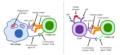 Activation of T and B cells.png 2,159 × 981; 263 KB
-
 Antibody Effector Mechanisms.png 5,025 × 4,043; 1.93 MB
-
 B cell (30702881796).jpg 1,633 × 2,401; 512 KB
-
 B cell activation naive to plasma cell.png 463 × 261; 98 KB
-
 B cell activation.png 651 × 1,000; 209 KB
-
 B cell function.png 813 × 280; 130 KB
-
 B cell signalling.png 960 × 720; 213 KB
-
 B-cell.png 500 × 375; 92 KB
-
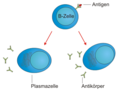 B-Zelle-Plasmazellen.png 453 × 342; 48 KB
-
 B8790rrt4.PNG 463 × 261; 104 KB
-
 BCelle.gif 512 × 473; 168 KB
-
 Bcr B-cell receptor.jpg 271 × 186; 10 KB
-
 Blausen 0624 Lymphocyte B cell (crop).png 1,165 × 1,130; 1.36 MB
-
 Blausen 0624 Lymphocyte B cell.png 1,500 × 1,500; 1.05 MB
-
 Clonal Deletion.png 1,440 × 816; 155 KB
-
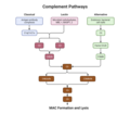 Complement Pathways.png 3,000 × 2,626; 371 KB
-
 CpG ODN effects.png 1,415 × 690; 83 KB
-
 Cèl·lules b de memòria.jpg 784 × 440; 56 KB
-
 Célula B (B Cell) (35795300934).jpg 1,176 × 1,872; 630 KB
-
 General Effector Mechanisms of B and T Cells.png 3,300 × 1,650; 554 KB
-
 Human B Lymphocyte (28942386960).jpg 2,624 × 2,936; 1.56 MB
-
 Human B Lymphocyte (29196367446).jpg 2,560 × 1,920; 605 KB
-
 Human B Lymphocyte - NIAID.jpg 2,560 × 1,920; 568 KB
-
 Human Cell Groups distributed by Cell Count and by Aggregate Cell Mass.jpg 3,162 × 2,096; 1.08 MB
-
 Humeral Secondary Immune Response.png 5,025 × 3,137; 1.06 MB
-
 Immunological Memory.png 5,025 × 4,579; 1.55 MB
-
 Model of Centroblast Development.png 960 × 540; 54 KB
-
 Sistèma immunitari - Activacion dei linfocits B dins l'òme.png 1,293 × 1,083; 201 KB
-
 T independent B cell Activation.png 4,844 × 2,018; 674 KB
-
 T-dependent B cell activation.png 960 × 720; 184 KB
-
 Transitional B cell development.PNG 303 × 217; 16 KB
-
 Vereenvoudigde weergave van het primaire immuunantwoord v2.png 1,948 × 1,534; 1.41 MB
-
 أنواع-خلايا-بي1.png 431 × 732; 225 KB
-
 خلية بي1.png 446 × 371; 145 KB
-
 ബി-ലസികാണുക്കളുടെ പ്രതിജനകബന്ധനവും ക്ലോണിക നിർധാരണവും.png 1,407 × 2,304; 1.4 MB


.jpg)


.png)


_(35795300934).jpg)

.jpg)
.jpg)


