Category:Biological diagrams in Russian
Jump to navigation
Jump to search
Media in category "Biological diagrams in Russian"
The following 121 files are in this category, out of 121 total.
-
 1414 Rods and Cones - ru.svg 2,075 × 3,112; 494 KB
-
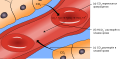 2325 Carbon Dioxide Transport - ru.svg 1,906 × 929; 35 KB
-
 Abdomen-head-thorax-ru.svg 793 × 452; 261 KB
-
 Abnormalsperm-ru.svg 369 × 529; 169 KB
-
 Adherens Junctions structural proteins-ru.svg 504 × 579; 185 KB
-
 Amino acid catabolism revised-ru.svg 905 × 636; 70 KB
-
 Amphibian brain ru.svg 198 × 159; 50 KB
-
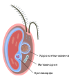
 Anatomy of an amiotic egg labeled ru.svg 8,934 × 4,520; 39 KB
-
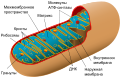 Animal mitochondrion diagram ru.svg 562 × 359; 216 KB
-
 Antagonist 2-ru.svg 694 × 468; 1 KB
-
 ApocritanAbdomen-ru.svg 800 × 700; 54 KB
-
 Apomorphy - Homoplasy-RU.svg 265 × 252; 30 KB
-
 Araneae ru.svg 744 × 524; 66 KB
-
 Archimollusc-ru.svg 702 × 512; 8.21 MB
-
 Average prokaryote cell- ru.svg 494 × 402; 116 KB
-
 Avocado seed diagram-ru.svg 720 × 500; 22 KB
-
 B6f subunit colored.jpg 1,522 × 1,200; 308 KB
-
 Bacteria Cytoskeleton-ru.svg 423 × 210; 17 KB
-
 Bacterial morphology diagram-ru.svg 825 × 762; 529 KB
-
 Biological and technological scales compared-ru.svg 1,062 × 755; 1.51 MB
-
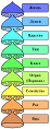 Biological classification L Pengo vflip ru.svg 230 × 590; 56 KB
-
 Biological classification L Pengo vflip russian.svg 230 × 590; 56 KB
-
 Biological classification L Pengo-ru.svg 230 × 590; 63 KB
-

-
 Blausen 0644 Mitochondria-ru.png 1,600 × 1,360; 1.69 MB
-
 Bufonidae male reproductive system ru.svg 1,000 × 1,000; 69 KB
-
 C2-фотосинтез.svg 512 × 419; 125 KB
-
 C4 and C3 temperature response curves - ru.svg 655 × 563; 15 KB
-
 C4 photosynthesis NAD-ME (ru).svg 910 × 549; 130 KB
-
 C4 photosynthesis NADP-ME type (ru).svg 910 × 311; 95 KB
-
 C4 photosynthesis PEPCK (ru).svg 910 × 549; 156 KB
-
 C4 plants(ru).svg 619 × 326; 34 KB
-
 Calvin-cycle4 ru.svg 836 × 766; 128 KB
-
 CAM cycle(ru).svg 1,020 × 488; 536 KB
-
 Catabolism schematic ru.svg 712 × 684; 2 KB
-
 Categoryweb 001.svg 1,488 × 1,098; 21 KB
-
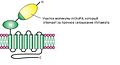
 Cell membrane detailed diagram 3-ru.svg 1,973 × 1,532; 517 KB
-
 Cellular tight junction-ru.svg 499 × 646; 142 KB
-
 Centrosome (standalone version)-ru.svg 3,280 × 3,160; 187 KB
-
 Centrum Morphology-ru.svg 565 × 453; 237 KB
-
 Characteristics of life-ru.svg 512 × 358; 18 KB
-
 Chiasma-ru.png 164 × 69; 4 KB
-
 ChIP-on-chip workflow overview-ru.svg 1,049 × 559; 20 KB
-
 Chlorosome.png 347 × 235; 13 KB
-
 Chromothripsis.png 800 × 493; 77 KB
-
 Citric acid cycle with aconitate 2 ru.svg 1,335 × 1,062; 635 KB
-
 Cladogram-example2-rus.gif 417 × 318; 3 KB
-
 CMVschema-ru.png 721 × 496; 82 KB
-
 CMVschema-ru.svg 721 × 496; 34 KB
-
 Cochlea-crosssection - ru.svg 512 × 433; 87 KB
-
 Complement pathway-ru.svg 743 × 881; 29 KB
-
 Complement-pathways-ru.svg 512 × 709; 63 KB
-
 Complete neuron cell diagram ru.svg 819 × 596; 570 KB
-
 Crispr - ru.png 2,100 × 1,700; 243 KB
-
 CRISPR overview - ru.svg 1,215 × 1,250; 211 KB
-
 CRISPR transfection - ru.svg 764 × 742; 257 KB
-
 CRISPR type I system - ru.svg 581 × 495; 149 KB
-
 CRISPR-Cas9 mode of action - ru.svg 1,551 × 442; 116 KB
-
 Cross section jellyfish ru.svg 860 × 451; 303 KB
-
 Cryptophyta-ru.svg 1,000 × 1,200; 164 KB
-
 Cumacea ru.svg 518 × 456; 360 KB
-
 Cyclic Photophosphorylation-ru.svg 476 × 178; 557 KB
-
 Cytochrome f interects plastocyanin.png 1,282 × 760; 122 KB
-
 Diagram human cell nucleus ru.svg 462 × 378; 112 KB
-
 Dinoflagellate genome - ru.svg 1,110 × 1,129; 301 KB
-
 DNA chemical structure RU.svg 1,500 × 1,750; 144 KB
-
 Dopamine pathways -ru.svg 479 × 335; 27 KB
-
 Dopamine serotonin rus.png 1,665 × 1,014; 463 KB
-
 Dromaeosaurus skull ru.svg 680 × 400; 112 KB
-
 Drupe fruit diagram-ru.svg 432 × 322; 31 KB
-
 ENaC.jpg 187 × 277; 20 KB
-
 Endomembrane system diagram ru.svg 612 × 486; 78 KB
-
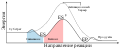 Enzyme catalysis energy levels 2-ru.svg 733 × 301; 2 KB
-
 First satge sbep2.png 1,024 × 270; 51 KB
-
 FtsZ cytokinesis ru.png 800 × 492; 73 KB
-
 FumaraseMechanismE1CB - ru.png 1,696 × 1,718; 78 KB
-
 Gap cell junction-ru.svg 582 × 409; 57 KB
-
 Grafic 4.JPG 2,379 × 1,430; 508 KB
-
 H pylori ulcer ru.jpg 800 × 603; 95 KB
-
 IDHcatalyticmechanism - ru.jpg 419 × 539; 62 KB
-
 Isopren emission of leaf-ru.svg 511 × 205; 1.98 MB
-
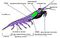 Krillanatomykils ru.jpg 592 × 380; 96 KB
-
 MGluR4.JPG 448 × 241; 14 KB
-
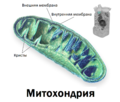
 Mitochondrion (standalone version)-ru.svg 3,280 × 3,160; 365 KB
-
 Neuron-rus.svg 460 × 319; 77 KB
-
 OxygenEvaluation.png 445 × 321; 64 KB
-
 Paracentric and pericentric inversions.png 326 × 256; 14 KB
-
 PEP Carboxylase Regulation(ru).png 1,299 × 739; 80 KB
-
 PFK-1 - ru.jpg 800 × 666; 248 KB
-
 Photoinhibition(ru).svg 676 × 494; 10 KB
-
 Photosynthese CO2-Konzentration (ru).svg 480 × 422; 27 KB
-
 Photosynthese Lichtmen(ru).svg 620 × 385; 55 KB
-
 Plant cell structure-ru-v1.png 900 × 740; 288 KB
-
 Plant cell structure-ru-v2.png 2,601 × 1,764; 920 KB
-
 Protone pomps.jpg 798 × 418; 109 KB
-
 PSI trimer.png 586 × 589; 347 KB
-
 PSI+LHC.png 787 × 1,173; 93 KB
-
 PSII-phosphorescent.png 810 × 1,040; 94 KB
-
 Puple bacteria anten.png 308 × 176; 10 KB
-
 PupleBacterPS.png 778 × 708; 25 KB
-
 Quadrum.jpg 723 × 473; 62 KB
-
 Reductive TCA cycle - ru.png 807 × 671; 65 KB
-
 Reversible phosphorylation of PPDK by PDRP(ru).svg 512 × 167; 59 KB
-
 RNAi-simplified-rus.png 701 × 599; 96 KB
-
 Scheme of axolotls' activity.jpg 2,328 × 1,680; 396 KB
-
 Sex determination-ru.jpg 490 × 363; 92 KB
-
 ShemaQ10.jpg 1,446 × 379; 68 KB
-
 SimpleCRISPR - ru.jpg 1,195 × 350; 68 KB
-
 Single-cell c4 photosynthesis(ru).svg 420 × 987; 43 KB
-
 SiRNA and RNAI rus.png 1,039 × 903; 38 KB
-
 Squalen epoxidation.PNG 3,024 × 875; 15 KB
-
 Tilacoid structure.png 748 × 548; 265 KB
-
 Translocation-4-20rus.png 464 × 345; 22 KB
-
 U curve.png 653 × 454; 34 KB
-
 WGCNA outline (russian).png 896 × 760; 276 KB
-
 Андрогеновый рецептор-7.png 1,280 × 960; 285 KB
-
 Взаимодействие фотосистем.png 968 × 802; 19 KB
-
 Мембрана хлоропласта.png 384 × 210; 43 KB
-
 Скрещивание.png 1,492 × 226; 17 KB
-
 Спектр действия фотосинтеза.svg 892 × 660; 20 KB
-
 Щелевой контакт.jpg 582 × 409; 147 KB


.svg)
.svg)
.svg)

.svg)


-ru.svg)


-ru.svg)


.png)

.svg)
.svg)
.svg)


.svg)


.png)


.svg)
.svg)