Category:Biomedical engineering
Jump to navigation
Jump to search
Main gallery: w:en:Biomedical engineering.
application of engineering principles and design concepts to medicine and biology | |||||
| Upload media | |||||
| Instance of |
| ||||
|---|---|---|---|---|---|
| Subclass of |
| ||||
| Said to be the same as | health technology, medical technology | ||||
| |||||
Subcategories
This category has the following 16 subcategories, out of 16 total.
Media in category "Biomedical engineering"
The following 128 files are in this category, out of 128 total.
-
 12 lead ECG.jpg 3,507 × 2,300; 1.59 MB
-
 3036810 13239 2010 27 Fig1 HTML.png 512 × 339; 52 KB
-
 3112189 pone.0020674.g001.png 512 × 278; 60 KB
-
 41392 2022 1024 Fig1 HTML.webp 1,799 × 1,357; 128 KB
-
-
 A comparison of blood pressure and photoplethysmogram signals.svg 800 × 400; 44 KB
-
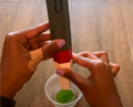
 A plasma expression.jpg 2,358 × 2,358; 982 KB
-

-

-
 AC microcapsule microphotographs.png 284 × 220; 101 KB
-
 Acaide505f01th.jpg 450 × 448; 64 KB
-
 Accessing vascular age from the photoplethysmogram.pdf 1,979 × 1,181; 697 KB
-
 Advanced medical imaging during noninjury ED visits.png 960 × 518; 32 KB
-
 Analysing photoplethysmogram signals.pdf 1,947 × 1,229; 513 KB
-
 Anatomisch 3D-model in Mimics.jpg 1,011 × 836; 90 KB
-
 ANEEB2017 v0.jpg 494 × 378; 24 KB
-
 AP microcapsule integrity, GI simulated transit.png 714 × 217; 196 KB
-
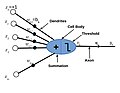 Artificialneuronhjdevias.jpeg 1,536 × 1,187; 192 KB
-
 Assessing effective connectivity in epileptogenic networks. A model-based simulation approach..pdf 1,239 × 1,754, 10 pages; 286 KB
-
 Atrioventricularvalve.jpg 400 × 320; 78 KB
-
 Autosyringe.png 1,023 × 825; 1,000 KB
-
 Biomedical and Biotechnology department at NIT Rourkela.jpg 3,264 × 2,448; 2.25 MB
-
 Biotechnology & Biomedical departments NITR.jpg 3,264 × 2,448; 2.26 MB
-
 Biotribology.jpeg 1,056 × 816; 151 KB
-
 BKG zaznam.jpg 901 × 623; 80 KB
-
 BMSEED’s electrophysiology module Intan 120-Channel.png 2,016 × 1,512; 2.52 MB
-
 Brain-computer interface (BCI) system.jpg 250 × 206; 10 KB
-
 Bulk vs surface erosion.jpg 382 × 215; 21 KB
-
 Cardiacoutput4.jpg 670 × 331; 56 KB
-
 Cardiacoutput7.jpg 500 × 498; 32 KB
-
 Cell capsule schematic.png 436 × 266; 49 KB
-
 Challenges and perspectives of organoid technologies.webp 1,500 × 1,387; 115 KB
-
 Circuito-Inibicao-Reciproca.tif 728 × 374; 20 KB
-
 Classes of photoplethysmogram (PPG) pulse wave shape.svg 1,200 × 250; 57 KB
-
 Clinical vs laboratory photoplethysmogram (PPG) pulse waves.svg 600 × 300; 36 KB
-
 Control drone by eeg.jpg 1,721 × 963; 363 KB
-
 Corticography recording.png 444 × 355; 427 KB
-
 Darwin XI's environment and experimental protocol..jpg 450 × 684; 143 KB
-
 Detecting atrial fibrillation (AF) from the photoplethysmogram (PPG).svg 700 × 800; 203 KB
-
 Dr. Amin Mahoutforoush.jpg 1,272 × 841; 372 KB
-
 Drugdeliverybrain.jpeg 1,440 × 1,113; 352 KB
-
 Electroretinographhjdeviass2.jpeg 2,884 × 1,409; 589 KB
-
 Estimating blood pressure from the photoplethysmogram.pdf 1,979 × 1,181; 382 KB
-
 Extracting respiratory signals from the photoplethysmogram (PPG).svg 576 × 288; 107 KB
-
-
 Finger vs ear photoplethysmogram (PPG) pulse waves.svg 600 × 300; 34 KB
-
 Flow cytometer structure.png 865 × 599; 1.98 MB
-
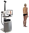 Formetric 4D 12x14cm RGB.jpg 1,417 × 1,627; 205 KB
-
 Graphical description of the Fick principle..jpg 500 × 210; 21 KB
-
 Heart Rate Display Using Arduino and LabView.png 1,334 × 750; 2.66 MB
-
 Hgmitralvalve.jpeg 1,440 × 1,113; 361 KB
-
 Hjdeviassarotic valve.jpeg 1,440 × 1,113; 496 KB
-

-
 Institut d'Investigacions Biomèdiques de Barcelona.JPG 1,536 × 2,048; 1.26 MB
-
 JindrichKopecekphoto2020.jpg 1,080 × 1,008; 257 KB
-
 Jude Odele - Makerere University.jpg 1,840 × 1,840; 121 KB
-
 Lower limb amputation levels.jpg 635 × 335; 49 KB
-
 Manually Stretching Microelectrode Array.jpg 1,920 × 1,080; 805 KB
-
 Material Models - Cardiovascular stent.jpg 600 × 400; 140 KB
-

-
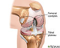 Medlineplustibial plateau.jpg 400 × 320; 69 KB
-
 Methodological advancement in brain organoid generation.webp 1,350 × 896; 124 KB
-
 MicroElectrode Array Stretching Simulating und Recording Equipment.jpg 3,024 × 4,032; 642 KB
-
 Micropear.png 3,233 × 2,425; 6.46 MB
-
 Modeling Respiratory system.png 600 × 600; 103 KB
-
 Modeling the Cardiovascular System.png 449 × 813; 54 KB
-
 ModeloArteriaGIF.gif 500 × 500; 72 KB
-
 Modelos con cuadrícula.png 650 × 220; 42 KB
-
 Modelos sin cuadrícula.png 520 × 220; 78 KB
-
 MRtractographyheartRat.png 512 × 395; 435 KB
-
 Myki project.jpg 8,466 × 7,018; 5 MB
-
 Myofiber architecture and MRtractography of heart.jpeg 512 × 395; 122 KB
-
 Neuromodulation targets for motor symptoms in Parkinson's disease.webp 2,052 × 1,085; 165 KB
-
 Neuronactivity.jpeg 1,440 × 1,113; 414 KB
-
 Neuroprosthetic technologies for sensorimotor disorders.webp 2,041 × 1,300; 319 KB
-
 Neurotrophic Electrode2.JPG 414 × 255; 8 KB
-
 Nonlinear analysis of the photoplethysmogram.svg 1,000 × 600; 209 KB
-
 Photoplethysmogram (PPG) pulse wave fiducial points.svg 304 × 800; 45 KB
-
 Photoplethysmogram (PPG) pulse wave indices.svg 304 × 800; 122 KB
-
 Photoplethysmogram (PPG) pulse wave.svg 500 × 330; 33 KB
-
 Photoplethysmogram pulse wave composition.svg 500 × 330; 39 KB
-
 Photoplethysmogram signal components.svg 1,000 × 470; 105 KB
-
 Pipeline for multiscale hyperdimensional analysis of organoids.webp 1,597 × 2,148; 661 KB
-
 Plateau of Tibia.jpeg 1,440 × 1,113; 500 KB
-
 Plethysmography.jpg 400 × 320; 55 KB
-

-
 Possible neuromodulation strategies for cognitive impairment and dementia.webp 2,048 × 1,182; 170 KB
-
 Projecte Barcino.jpg 775 × 537; 125 KB
-
 Pulse contour analysis..jpg 501 × 587; 45 KB
-
 Pwv measurement fig.svg 1,600 × 450; 352 KB
-
 Recording single-lead ECG using a Withings Move ECG watch.jpg 1,471 × 1,591; 367 KB
-
 Reflexo H.gif 488 × 279; 9 KB
-
 Respiratory modulations of the photoplethysmogram.svg 691 × 288; 109 KB
-
 Ryan O'Shea Northstar Photo.JPG 5,472 × 3,648; 6.86 MB
-
 Sarcomerelengthtension.png 512 × 338; 88 KB
-
-
-
-
-
-
-
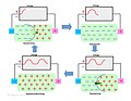
 Schematic of different strategies to vascularization of brain organoid.webp 1,350 × 874; 138 KB
-
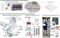
 Schematic representation of the biomedical applications of microswimmers.webp 3,365 × 2,200; 616 KB
-
 Science edunihgovinfo fig02.gif 500 × 292; 11 KB
-
 Short photoplethysmogram (PPG) signal.svg 1,000 × 470; 31 KB
-
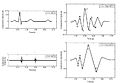
 Simulated photoplethysmogram pulse waves.png 2,999 × 1,533; 599 KB
-
 Stretchable Microelectrode Array (sMEA) before and during stretch.png 2,205 × 1,404; 3.42 MB
-
 Stretchable Microelectrode Array (sMEA) with PDMS glue.jpg 3,024 × 4,032; 1.7 MB
-
 Stretchable Microelectrode Array (sMEA) with white glue.jpg 2,732 × 2,732; 1.31 MB
-
 Surface and Bulk Erosion.jpg 241 × 551; 9 KB
-
 The concept of neuroregeneration.png 512 × 611; 225 KB
-

-
 The neuroprosthetic system..gif 400 × 131; 5 KB
-
 Tibia-CT-Biomechanics-part1.png 990 × 814; 362 KB
-
 Towards large-scale bidirectional neuroprostheses for memory disorders.webp 2,044 × 1,615; 256 KB
-
-
-
-
 Triggering a single-lead ECG recording on a Withings Move ECG watch.jpg 1,249 × 1,544; 319 KB
-
 TxBDC Logo.png 413 × 122; 4 KB
-
 Variations of the velocity across the tricuspid valve.png 512 × 384; 277 KB
-
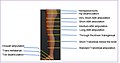
 Volume conductor model.png 466 × 327; 261 KB
-
 VolumeConductorprinciple.jpeg 1,536 × 1,187; 226 KB
-
 Wavevelocity.png 468 × 350; 53 KB
-
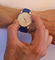
 Withings Move ECG watch worn on wrist.jpg 1,003 × 1,003; 198 KB
-
 Wrist pulse oximeter.jpg 1,347 × 726; 75 KB
-
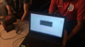
 צילום כיתת ביורפואה ומערכות רפואיות בבית חינוך ירקון.jpg 1,920 × 1,080; 143 KB
-
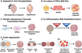
 “Mini-brains” generated from PSCs.webp 1,971 × 465; 276 KB


_system.jpg)


_pulse_waves.svg)


_from_the_photoplethysmogram_(PPG).svg)


.svg)
_and_photoplethysmogram_(PPG).png)
_pulse_waves.svg)


_pulse_wave.svg)


_signal.svg)

_before_and_during_stretch.png)
_with_PDMS_glue.jpg)
_with_white_glue.jpg)


_pulse_wave_shape.svg)
_pulse_wave_fiducial_points.svg)
_pulse_wave_indices.svg)
_images_from_a_Parkinson%27s_patient_before_and_after_fetal_tissue_transplantation..jpg)