Category:CT images of osteoma
Jump to navigation
Jump to search
Media in category "CT images of osteoma"
The following 29 files are in this category, out of 29 total.
-
 Osteom - Siebbeinzellen.jpg 844 × 484; 30 KB
-
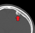
 Osteom der Kalotte links okzipital 46W - CT - 001 - Annotation.jpg 1,592 × 1,095; 206 KB
-
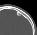
 Osteom der Kalotte links okzipital 46W - CT - 001.jpg 1,592 × 1,095; 209 KB
-
 Osteom der Kieferhoehle rechts 65M - CT axial - 001 - Annotation.jpg 1,108 × 1,362; 95 KB
-
 Osteom der Kieferhoehle rechts 65M - CT axial - 001.jpg 1,108 × 1,362; 83 KB
-
 Osteom der Scapula 87W - CT - 001.jpg 2,033 × 1,027; 155 KB
-
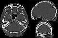
 Osteom der Schaedelkalotte 75W - CT - 001.jpg 1,473 × 1,361; 223 KB
-
 Osteom der Schaedelkalotte rechts 75W - CT - 001.jpg 3,050 × 1,135; 463 KB
-
 Osteom der Stirnhoehle CT.jpg 500 × 606; 57 KB
-
 Osteom der Stirnhoehle rechts 48W - CT - 001.jpg 2,012 × 1,319; 241 KB
-
 Osteom im Os ilium 69W - CT und CR - 001 - Annotation.jpg 2,691 × 773; 244 KB
-
 Osteom im Os ilium 69W - CT und CR - 001.jpg 2,691 × 773; 320 KB
-
 Osteom im Sinus frontalis rechts 84M - CT - 001 - Annotation.jpg 1,418 × 985; 185 KB
-
 Osteom im Sinus frontalis rechts 84M - CT - 001.jpg 1,418 × 985; 230 KB
-
 Osteom in den Ethmoidalzellen links 80W - CT axial - 001 - Annotation.jpg 1,489 × 1,683; 220 KB
-
 Osteom in den Ethmoidalzellen links 80W - CT axial - 001.jpg 1,489 × 1,683; 271 KB
-
 Osteom Os capitatum 30M - CT - 001.jpg 1,911 × 565; 122 KB
-
 Osteom Stirnhoehle CT KF axial 001.png 914 × 1,008; 244 KB
-
 Osteom Stirnhoehle CT KF axial 002.png 830 × 452; 107 KB
-
 Osteoma.png 962 × 925; 209 KB
-
 OsteomaMark.png 962 × 925; 208 KB
-
 Solides Osteom der Stirnhoehle rechts 79M - CT - 001 - Annotation.jpg 2,036 × 1,234; 279 KB
-
 Solides Osteom der Stirnhoehle rechts 79M - CT - 001.jpg 2,036 × 1,234; 308 KB
-
 Typischer Befund eines Osteoms im Os ilium 72W - CT und CR - 001 - Annotation.jpg 1,779 × 1,387; 290 KB
-
 Typischer Befund eines Osteoms im Os ilium 72W - CT und CR - 001.jpg 1,779 × 1,387; 291 KB
-
 Typisches Osteom im distalen Radius 26M - CT und CR - 001 - Annotation.jpg 1,486 × 1,111; 167 KB
-
 Typisches Osteom im distalen Radius 26M - CT und CR - 001.jpg 1,486 × 1,111; 163 KB
-
 Typisches solides Osteom der Schaedelkalotte 75W - CT - 001.jpg 2,791 × 1,408; 379 KB
-
 Typisches solides Osteom der Schaedelkalotte 75W - CT axial und VR - 001.jpg 2,363 × 1,320; 413 KB


