Category:Cell adhesion
Zur Navigation springen
Zur Suche springen
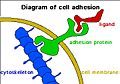
attachment of a cell, to another cell or to an underlying substrate, by cell adhesion molecules | |||||
| Medium hochladen | |||||
| Ist ein(e) | |||||
|---|---|---|---|---|---|
| Unterklasse von | |||||
| |||||
Unterkategorien
Es werden 7 von insgesamt 7 Unterkategorien in dieser Kategorie angezeigt:
In Klammern die Anzahl der enthaltenen Kategorien (K), Seiten (S), Dateien (D)
Medien in der Kategorie „Cell adhesion“
Folgende 200 Dateien sind in dieser Kategorie, von 1.115 insgesamt.
(vorherige Seite) (nächste Seite)-
-Adrenergic-Inhibition-of-Contractility-in-L6-Skeletal-Muscle-Cells-pone.0022304.s002.ogv 17 s, 320 × 240; 877 KB
-
-
-
A-Comprehensive-Panel-of-Three-Dimensional-Models-for-Studies-of-Prostate-Cancer-Growth-Invasion-pone.0010431.s013.ogv 1 min 6 s, 524 × 384; 8,71 MB
-
A-Gene-Expression-Signature-of-Invasive-Potential-in-Metastatic-Melanoma-Cells-pone.0008461.s008.ogv 15 s, 640 × 480; 2,11 MB
-
A-Gene-Expression-Signature-of-Invasive-Potential-in-Metastatic-Melanoma-Cells-pone.0008461.s009.ogv 15 s, 640 × 480; 2,37 MB
-
A-Mathematical-Model-for-EphEphrin-Directed-Segregation-of-Intermingled-Cells-pone.0111803.s003.ogv 1 min 12 s, 640 × 480; 19,37 MB
-
A-Mathematical-Model-for-EphEphrin-Directed-Segregation-of-Intermingled-Cells-pone.0111803.s004.ogv 1 min 36 s, 640 × 480; 59,23 MB
-
A-Multi-cell-Multi-scale-Model-of-Vertebrate-Segmentation-and-Somite-Formation-pcbi.1002155.s016.ogv 1 min 3 s, 640 × 480; 6,71 MB
-
A-Multi-cell-Multi-scale-Model-of-Vertebrate-Segmentation-and-Somite-Formation-pcbi.1002155.s017.ogv 1 min 2 s, 640 × 480; 6,06 MB
-
A-Multi-cell-Multi-scale-Model-of-Vertebrate-Segmentation-and-Somite-Formation-pcbi.1002155.s018.ogv 1 min 2 s, 640 × 480; 6,04 MB
-
-
A-Multi-cell-Multi-scale-Model-of-Vertebrate-Segmentation-and-Somite-Formation-pcbi.1002155.s020.ogv 2 min 5 s, 640 × 480; 5,94 MB
-
-
-
-
-
-
-
-
-
-
-
-
-
-
-
-
-
-
-
-
-
-
-
-
-
A-Role-for-PP1NIPP1-in-Steering-Migration-of-Human-Cancer-Cells-pone.0040769.s002.ogv 7,2 s, 696 × 520; 237 KB
-
A-Role-for-PP1NIPP1-in-Steering-Migration-of-Human-Cancer-Cells-pone.0040769.s003.ogv 7,2 s, 696 × 520; 226 KB
-
A-Role-for-PP1NIPP1-in-Steering-Migration-of-Human-Cancer-Cells-pone.0040769.s004.ogv 1,8 s, 696 × 520; 231 KB
-
A-Role-for-PP1NIPP1-in-Steering-Migration-of-Human-Cancer-Cells-pone.0040769.s005.ogv 3,6 s, 696 × 520; 286 KB
-
A-Role-for-PP1NIPP1-in-Steering-Migration-of-Human-Cancer-Cells-pone.0040769.s006.ogv 5,1 s, 696 × 520; 208 KB
-
A-Role-for-PP1NIPP1-in-Steering-Migration-of-Human-Cancer-Cells-pone.0040769.s007.ogv 5,1 s, 696 × 520; 155 KB
-
A-Role-for-PP1NIPP1-in-Steering-Migration-of-Human-Cancer-Cells-pone.0040769.s008.ogv 3,6 s, 696 × 520; 222 KB
-
A-Role-for-PP1NIPP1-in-Steering-Migration-of-Human-Cancer-Cells-pone.0040769.s009.ogv 9,0 s, 696 × 520; 263 KB
-
A-Role-for-PP1NIPP1-in-Steering-Migration-of-Human-Cancer-Cells-pone.0040769.s010.ogv 7,0 s, 696 × 520; 371 KB
-
A-Role-for-PP1NIPP1-in-Steering-Migration-of-Human-Cancer-Cells-pone.0040769.s011.ogv 7,2 s, 696 × 520; 434 KB
-
A-Role-for-PP1NIPP1-in-Steering-Migration-of-Human-Cancer-Cells-pone.0040769.s012.ogv 3,6 s, 696 × 520; 252 KB
-
A-Role-for-PP1NIPP1-in-Steering-Migration-of-Human-Cancer-Cells-pone.0040769.s013.ogv 3,6 s, 696 × 520; 309 KB
-
A-Role-for-PP1NIPP1-in-Steering-Migration-of-Human-Cancer-Cells-pone.0040769.s014.ogv 3,6 s, 696 × 520; 125 KB
-
A-Role-for-PP1NIPP1-in-Steering-Migration-of-Human-Cancer-Cells-pone.0040769.s015.ogv 3,6 s, 696 × 520; 225 KB
-
-
-
-
-
-
-
-
-
-
-
-
-
-
-
-
-
-
-
-
-
-
-
-
-
-
-
-
-
-
-
 Adhesion diagram.jpg 359 × 252; 84 KB
-
Adhesion-Failures-Determine-the-Pattern-of-Choroidal-Neovascularization-in-the-Eye-A-Computer-pcbi.1002440.s017.ogv 1 min 1 s, 637 × 616; 7,11 MB
-
Adhesion-Failures-Determine-the-Pattern-of-Choroidal-Neovascularization-in-the-Eye-A-Computer-pcbi.1002440.s018.ogv 1 min 1 s, 588 × 568; 6,38 MB
-
-
Adhesion-Failures-Determine-the-Pattern-of-Choroidal-Neovascularization-in-the-Eye-A-Computer-pcbi.1002440.s020.ogv 1 min 1 s, 588 × 568; 6,55 MB
-
Adhesion-Failures-Determine-the-Pattern-of-Choroidal-Neovascularization-in-the-Eye-A-Computer-pcbi.1002440.s021.ogv 1 min 1 s, 637 × 616; 7,29 MB
-
Adhesion-Failures-Determine-the-Pattern-of-Choroidal-Neovascularization-in-the-Eye-A-Computer-pcbi.1002440.s022.ogv 1 min 1 s, 637 × 616; 7,16 MB
-
-
-
-
-
-
-
-
-
-
-
-
-
-
-
-
-
-
-
-
-
-
-
-
-
-
-
-
-
-
-
-
-
-
-
-
-
An-Analysis-of-Trafficking-Receptors-Shows-that-CD44-and-P-Selectin-Glycoprotein-Ligand-1-Video 1.ogv 12 s, 1.222 × 866; 536 KB
-
An-Analysis-of-Trafficking-Receptors-Shows-that-CD44-and-P-Selectin-Glycoprotein-Ligand-1-Video 2.ogv 12 s, 1.222 × 731; 424 KB
-
An-Analysis-of-Trafficking-Receptors-Shows-that-CD44-and-P-Selectin-Glycoprotein-Ligand-1-Video 3.ogv 1 min 19 s, 800 × 600; 9,23 MB
-
An-Analysis-of-Trafficking-Receptors-Shows-that-CD44-and-P-Selectin-Glycoprotein-Ligand-1-Video 4.ogv 1 min 20 s, 800 × 600; 7,37 MB
-
An-Analysis-of-Trafficking-Receptors-Shows-that-CD44-and-P-Selectin-Glycoprotein-Ligand-1-Video 5.ogv 1 min 23 s, 800 × 600; 8,75 MB
-
An-Analysis-of-Trafficking-Receptors-Shows-that-CD44-and-P-Selectin-Glycoprotein-Ligand-1-Video 6.ogv 1 min 19 s, 800 × 600; 6,18 MB
-
An-Analysis-of-Trafficking-Receptors-Shows-that-CD44-and-P-Selectin-Glycoprotein-Ligand-1-Video 7.ogv 1 min 20 s, 800 × 600; 6,51 MB
-
-
An-Improved-Chamber-for-Direct-Visualisation-of-Chemotaxis-pone.0015309.s001.ogv 12 s, 316 × 538; 2,13 MB
-
An-Improved-Chamber-for-Direct-Visualisation-of-Chemotaxis-pone.0015309.s002.ogv 12 s, 316 × 531; 2,29 MB
-
-
-
-
-
-
-
-
-
-
-
Arp23-complex-activity-in-filopodia-of-spreading-cells-1471-2121-9-65-S1.ogv 1 min 5 s, 434 × 440; 1,65 MB
-
Arp23-complex-activity-in-filopodia-of-spreading-cells-1471-2121-9-65-S4.ogv 3,8 s, 210 × 288; 152 KB
-
Arp23-complex-activity-in-filopodia-of-spreading-cells-1471-2121-9-65-S5.ogv 20 s, 750 × 666; 418 KB
-
Arp23-complex-activity-in-filopodia-of-spreading-cells-1471-2121-9-65-S6.ogv 21 s, 614 × 394; 2,49 MB
-
-
-
-
-
-
-
-
-
-
-
-
-
-
-
-
-
-
-
-
-
-
Assembly-of-the-Murine-Leukemia-Virus-Is-Directed-towards-Sites-of-Cell–Cell-Contact-pbio.1000163.s009.ogv 40 s, 1.024 × 512; 3,32 MB
-
Assembly-of-the-Murine-Leukemia-Virus-Is-Directed-towards-Sites-of-Cell–Cell-Contact-pbio.1000163.s010.ogv 16 s, 1.024 × 512; 4,15 MB
-
Assembly-of-the-Murine-Leukemia-Virus-Is-Directed-towards-Sites-of-Cell–Cell-Contact-pbio.1000163.s011.ogv 11 s, 1.024 × 512; 6,81 MB
-
Assembly-of-the-Murine-Leukemia-Virus-Is-Directed-towards-Sites-of-Cell–Cell-Contact-pbio.1000163.s012.ogv 5,6 s, 1.024 × 512; 2,06 MB
-
Assembly-of-the-Murine-Leukemia-Virus-Is-Directed-towards-Sites-of-Cell–Cell-Contact-pbio.1000163.s013.ogv 16 s, 1.024 × 512; 2,32 MB
-
-
Assembly-of-the-Murine-Leukemia-Virus-Is-Directed-towards-Sites-of-Cell–Cell-Contact-pbio.1000163.s015.ogv 9,9 s, 1.024 × 512; 3,1 MB
-
Assembly-of-the-Murine-Leukemia-Virus-Is-Directed-towards-Sites-of-Cell–Cell-Contact-pbio.1000163.s016.ogv 8,8 s, 1.024 × 512; 1,91 MB
-
-
Assembly-of-the-Murine-Leukemia-Virus-Is-Directed-towards-Sites-of-Cell–Cell-Contact-pbio.1000163.s018.ogv 45 s, 1.077 × 404; 18,19 MB
-
-
-
-
-
-
-
Biological-soliton-in-multicellular-movement-srep02272-s1.ogv 28 s, 1.365 × 1.024; 5,68 MB
-
Biological-soliton-in-multicellular-movement-srep02272-s10.ogv 15 s, 1.280 × 720; 1,1 MB
-
Biological-soliton-in-multicellular-movement-srep02272-s11.ogv 15 s, 1.280 × 720; 6,04 MB
-
Biological-soliton-in-multicellular-movement-srep02272-s12.ogv 6,0 s, 1.280 × 720; 5,65 MB
-
Biological-soliton-in-multicellular-movement-srep02272-s13.ogv 6,0 s, 1.280 × 720; 802 KB
-
Biological-soliton-in-multicellular-movement-srep02272-s14.ogv 1 min 12 s, 640 × 480; 6,14 MB
-
Biological-soliton-in-multicellular-movement-srep02272-s2.ogv 36 s, 640 × 480; 3,84 MB
-
Biological-soliton-in-multicellular-movement-srep02272-s3.ogv 36 s, 640 × 480; 5,77 MB
-
Biological-soliton-in-multicellular-movement-srep02272-s4.ogv 18 s, 1.365 × 1.024; 5,4 MB
-
Biological-soliton-in-multicellular-movement-srep02272-s5.ogv 36 s, 1.365 × 1.024; 6,13 MB
-
Biological-soliton-in-multicellular-movement-srep02272-s6.ogv 36 s, 1.365 × 1.024; 4,29 MB
-
Biological-soliton-in-multicellular-movement-srep02272-s7.ogv 24 s, 1.365 × 1.024; 8,79 MB
-
Biological-soliton-in-multicellular-movement-srep02272-s8.ogv 1 min 12 s, 640 × 480; 15,54 MB
-
Biological-soliton-in-multicellular-movement-srep02272-s9.ogv 15 s, 1.280 × 720; 10,4 MB
-
