Category:Central sulcus
Jump to navigation
Jump to search
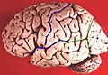
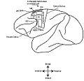
prominent landmark of the brain (primary motor cortex), separating the parietal lobe from the frontal lobe and the primary motor cortex from the primary somatosensory cortex. | |||||
| Upload media | |||||
| Instance of |
| ||||
|---|---|---|---|---|---|
| Subclass of |
| ||||
| Named after | |||||
| Different from | |||||
| |||||
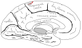
Central sulcus (the fissure of Rolando, the Rolandic fissure).
Media in category "Central sulcus"
The following 42 files are in this category, out of 42 total.
-
 Blausen 0103 Brain Sensory&Motor ar.png 1,600 × 960; 5.87 MB
-
 Blausen 0103 Brain Sensory&Motor.png 1,600 × 960; 993 KB
-
 Brodmann areas of human lateral frontal cortex.png 1,891 × 889; 2.31 MB
-
 Cambridge Natural History Mammalia Fig 273.png 532 × 374; 33 KB
-
 Central sulcus animation small.gif 150 × 150; 586 KB
-
 Central sulcus animation.gif 300 × 300; 1.75 MB
-
 Central sulcus diagram.png 300 × 190; 27 KB
-
 Central sulcus superior view.png 363 × 366; 134 KB
-
 Central sulcus.png 324 × 209; 27 KB
-
Cerebral Hemisphere Demonstration - Sanjoy Sanyal - Neuroscience Lab Fall 2013 1 (from 1m24s to 2m36s) Central sulcus.webm 1 min 12 s, 852 × 480; 12.71 MB
-
 Cunningham cerebral sulci.png 1,252 × 790; 2.84 MB
-
 Dorsal-cortex front.png 471 × 465; 259 KB
-
 Einstein brain.jpg 1,022 × 348; 139 KB
-
 Figure3 3-es.png 813 × 615; 358 KB
-
 Figure3 3.jpg 813 × 615; 307 KB
-
 Five sulci measured.png 1,660 × 1,215; 2.36 MB
-
 FrontalCapts.png 2,309 × 911; 927 KB
-
 FrontalCaptsLateral.png 1,252 × 829; 558 KB
-
 Gray1198.png 446 × 350; 24 KB
-
 Gray725 central sulcus.png 255 × 600; 63 KB
-
 Gray726 central sulcus.svg 992 × 573; 143 KB
-
 Gray727 central sulcus.svg 1,025 × 598; 18 KB
-
 Human brain lateral view description.JPG 701 × 487; 49 KB
-
 Human motor cortex topography.png 869 × 551; 32 KB
-

-
 Lateral aspect of left hemispheres showing main sulci.jpg 355 × 860; 275 KB
-
 Lobes.png 909 × 671; 382 KB
-
 LobesCaptsLateral.png 1,201 × 710; 441 KB
-
 LobesCaptsMedial1.png 1,063 × 755; 470 KB
-
 Macaque monkey's premotor areas.jpg 1,200 × 1,108; 156 KB
-
 Motor Cortex monkey.jpg 1,233 × 1,194; 147 KB
-
 ParietCapts lateral.png 1,139 × 758; 407 KB
-
 ParietCapts.png 2,337 × 878; 819 KB
-

-
 Pre- and post-central gyrus, right hemisphere cropped.png 426 × 488; 140 KB
-
 Pre- and post-central gyrus, right hemisphere.jpg 884 × 1,394; 1.13 MB
-
 Sillon occipital ant.png 669 × 519; 122 KB
-
 Sillons vue externe.png 888 × 492; 361 KB
-
 Sulcus centralis - Identification axial - MRI T2.jpg 681 × 795; 69 KB
-
 Sulcus centralis - Identification sagittal - MRI T2.jpg 912 × 700; 66 KB
-
 Superficial anatomy of the inferior parietal lobule (IPL).png 454 × 390; 349 KB
-
 Thickness of an humn adult cerebral cortex.jpg 926 × 284; 148 KB


.png)