Category:Chitin
Jump to navigation
Jump to search
long-chain polymer of a N-acetylglucosamine  | |||||
| Upload media | |||||
| Instance of | |||||
|---|---|---|---|---|---|
| Part of |
| ||||
| Has part(s) | |||||
| Mass |
| ||||
| |||||
Media in category "Chitin"
The following 41 files are in this category, out of 41 total.
-
 02 03 chitin, Fungi (M. Piepenbring).png 3,070 × 2,302; 312 KB
-
 1,6D-Glucan-Melanin-Chitin- Komplex als Hohlfaser.tif 568 × 642; 921 KB
-
 1,6D-Glucan-Melanin-Chitin-Komplex - die Hohlfaser..jpg 349 × 277; 21 KB
-
 Abb1.11 Fungi Strukturformeln Chitin Ergosterol 2021 (M. Piepenbring).png 3,000 × 2,250; 277 KB
-
 Alpha-Chitin large.gif 1,120 × 1,217; 134 KB
-
 Alpha-Chitin mini.gif 410 × 426; 27 KB
-
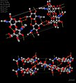 Alpha-Chitinlarge.jpg 1,120 × 1,217; 114 KB
-
 Chitin and Chitosan.jpg 890 × 1,069; 134 KB
-
 Chitin and chitosan.jpg 675 × 340; 45 KB
-
 Chitin Chitosan.svg 795 × 154; 80 KB
-
 Chitin fixed.png 353 × 230; 6 KB
-
 Chitin glucose and cellulose.svg 512 × 274; 290 KB
-
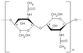
 Chitin Haworth.svg 437 × 244; 32 KB
-
 Chitin immune response.svg 482 × 313; 64 KB
-
 Chitin molecule.jpg 443 × 281; 40 KB
-
 Chitin structure.png 402 × 344; 6 KB
-
 Chitin.png 353 × 230; 2 KB
-
 Chitin.svg 512 × 302; 13 KB
-
 Chitine.png 348 × 265; 4 KB
-
 Chitobiose.svg 361 × 234; 32 KB
-
 Electrospray von Chtin gelöst in ionischer Flüssigkeit.jpg 2,048 × 1,536; 1.83 MB
-
 Exochitinase.png 1,908 × 504; 110 KB
-
 Glanzkaefer.jpg 800 × 600; 188 KB
-
 Haworth projection of chitin.svg 512 × 360; 7 KB
-
 High-resolution transmission electron microscopy image of chitin.jpg 732 × 323; 132 KB
-
Micro-CT-Imaging-of-Denatured-Chitin-by-Silver-to-Explore-Honey-Bee-and-Insect-Pathologies-pone.0027448.s002.ogv 6.7 s, 1,392 × 784; 2.91 MB
-
Micro-CT-Imaging-of-Denatured-Chitin-by-Silver-to-Explore-Honey-Bee-and-Insect-Pathologies-pone.0027448.s003.ogv 6.7 s, 1,392 × 784; 3.41 MB
-
Micro-CT-Imaging-of-Denatured-Chitin-by-Silver-to-Explore-Honey-Bee-and-Insect-Pathologies-pone.0027448.s004.ogv 6.7 s, 1,392 × 784; 3.01 MB
-
Micro-CT-Imaging-of-Denatured-Chitin-by-Silver-to-Explore-Honey-Bee-and-Insect-Pathologies-pone.0027448.s005.ogv 6.7 s, 1,392 × 784; 3.49 MB
-
Micro-CT-Imaging-of-Denatured-Chitin-by-Silver-to-Explore-Honey-Bee-and-Insect-Pathologies-pone.0027448.s006.ogv 6.7 s, 1,392 × 784; 4.54 MB
-
Micro-CT-Imaging-of-Denatured-Chitin-by-Silver-to-Explore-Honey-Bee-and-Insect-Pathologies-pone.0027448.s007.ogv 6.7 s, 1,392 × 784; 4.35 MB
-
Micro-CT-Imaging-of-Denatured-Chitin-by-Silver-to-Explore-Honey-Bee-and-Insect-Pathologies-pone.0027448.s008.ogv 6.7 s, 1,392 × 784; 3.84 MB
-
Micro-CT-Imaging-of-Denatured-Chitin-by-Silver-to-Explore-Honey-Bee-and-Insect-Pathologies-pone.0027448.s009.ogv 6.7 s, 1,392 × 784; 4.26 MB
-
 Nanostructural Organization of Naturally Occurring Composites Part I.pdf 1,250 × 1,650, 9 pages; 5.17 MB
-
 Nanostructural Organization of Naturally Occurring Composites Part II.pdf 1,250 × 1,650, 8 pages; 4.05 MB
-
 Preparation of NC and BC films.png 1,325 × 948; 169 KB
-
 Production of BCNF-CNF films.png 1,724 × 1,044; 196 KB
-

-
 QUITINA.gif 853 × 192; 5 KB
-
 QUITINA.svg 853 × 192; 30 KB
-
 Structure of Chitin.png 570 × 212; 41 KB
.png)

.png)


