Category:Ectoderm
Jump to navigation
Jump to search
germ layer that forms the brain, spinal cord, epidermis and more | |||||
| Upload media | |||||
| Instance of |
| ||||
|---|---|---|---|---|---|
| Subclass of |
| ||||
| Different from | |||||
| |||||
Media in category "Ectoderm"
The following 148 files are in this category, out of 148 total.
-
 2912 Neurulation-02 nltxt.jpg 2,500 × 2,500; 2.1 MB
-
 41392 2022 1024 Fig1 HTML.webp 1,799 × 1,357; 128 KB
-

-

-
-
 Abatus cordatus Long-spined juvenile (J2) (01).jpg 1,032 × 1,402; 1.43 MB
-
 Abatus cordatus Long-spined juvenile (J2).jpg 879 × 1,080; 781 KB
-
 Abatus cordatus Post-gastrular stage.jpg 930 × 1,284; 1.04 MB
-
 AER Apikale ektodermale Randleiste in der Extremitätenknospe.jpg 960 × 720; 131 KB
-
-

-
 Amphioxus Origin of the mesoderm.jpg 1,802 × 756; 793 KB
-

-
-
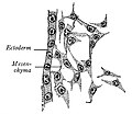 Avea Mesenchyma from the head of a thirty-six-hour chick embryo..jpg 833 × 723; 239 KB
-
-
-
-

-
-

-

-
-
-
-
-
-

-
 Ciliary marginal zone in vertebrates.jpg 1,543 × 668; 294 KB
-
 Clytia hemisphaerica day2 planula.jpg 1,590 × 1,590; 2.97 MB
-
 Coelomate 01.png 250 × 250; 12 KB
-
-
 Cortical neurogenesis in the mouse embryo.png 1,143 × 815; 530 KB
-
 Cresta neural.jpg 1,300 × 888; 85 KB
-
 Crucial biomolecules expression in an embryonic mouse at 9.5 days.jpg 797 × 883; 427 KB
-
 Development of the notochord in CS9 to CS12 (19–30 days).tiff 1,300 × 1,308; 1.82 MB
-

-

-
 Different degrees of EMT correlate with different tissue morphologies a.jpg 968 × 1,292; 576 KB
-
 Differentiation of the mesoderm in holoblastic and meroblastic types of development.jpg 1,617 × 1,467; 1,022 KB
-
 Differentiation oi Zygote and Cells (Hypothetical).png 565 × 605; 538 KB
-
 Diversity of vertebrate gastrulation.jpg 1,073 × 1,021; 415 KB
-
 Early gastrulation in amphibian embryos.png 3,570 × 1,358; 1,007 KB
-
 Early human embryo (01).jpg 675 × 563; 209 KB
-
 Early human embryo (02).jpg 915 × 432; 257 KB
-
 Early human embryo.jpg 761 × 738; 272 KB
-
 EB1911 Hydromedusae - Budding from the Ectoderm in Margellium.jpg 869 × 667; 143 KB
-
 EB1911 Lamellibranchia - development of Ostrea edulis.jpg 775 × 985; 261 KB
-
 EB1911 Peripatus - Series of Embryos.jpg 1,011 × 723; 218 KB
-
 Ectoderm-ar.png 274 × 176; 41 KB
-
 Ectopleura crocea. blastula (left) morula (right).jpg 1,074 × 825; 557 KB
-
 Embryonic and extraembryonic ectoderm demarcation in the amniochorionic fold.jpg 1,200 × 1,937; 1.62 MB
-
 Embryonic development in mice versus primates.jpg 1,016 × 1,286; 798 KB
-
-
-
-
-
-
-
-
-
 Epithelial differentiation is delayed in Bmp7 null endoderm.jpg 1,010 × 801; 666 KB
-
 Experimental manipulation of the gastrulation mode in different organisms.jpg 1,166 × 1,480; 850 KB
-

-

-
-
 Formation and patterning of the mouse neural tube.png 1,305 × 1,505; 636 KB
-
 Formation of the primitive body plan following gastrulation in the mouse.png 1,279 × 1,187; 1,016 KB
-
 Four diagrams showing hypothetical stages of early human embryos.jpg 1,631 × 1,434; 943 KB
-
 Frog embryo Sagittal section (2).jpg 782 × 694; 424 KB
-
 Fundulus heteroclitus presumptive organ-forming areas of the blastoderm.jpg 1,020 × 796; 779 KB
-
 Further Differentiation of Zygote (Hypothetical).png 552 × 445; 422 KB
-

-
 Gray29.png 500 × 306; 15 KB
-
 Histological sections of three stage 8 (17–19 days) human embryos.tiff 1,234 × 652; 702 KB
-
 Human embryo Section of embryonic rudiment in Peters' ovum (first week).jpg 1,141 × 857; 540 KB
-

-
-
Integrin-α5-Coordinates-Assembly-of-Posterior-Cranial-Placodes-in-Zebrafish-and-Enhances-Fgf-pone.0027778.s007.ogv 5.4 s, 1,388 × 1,040; 1.07 MB
-
Integrin-α5-Coordinates-Assembly-of-Posterior-Cranial-Placodes-in-Zebrafish-and-Enhances-Fgf-pone.0027778.s008.ogv 4.9 s, 1,388 × 1,040; 327 KB
-
Integrin-α5-Coordinates-Assembly-of-Posterior-Cranial-Placodes-in-Zebrafish-and-Enhances-Fgf-pone.0027778.s009.ogv 5.0 s, 1,388 × 1,040; 408 KB
-
 Kirkes' handbook of physiology (1907) (14769687492).jpg 1,091 × 918; 435 KB
-

-
 Li 46 green (antibody) 2-18.jpg 200 × 191; 27 KB
-
 Limb bud diagram.jpg 858 × 1,471; 204 KB
-

-
 Lumbricus Segmentation and early stages of development.jpg 1,124 × 1,088; 457 KB
-
-

-
 Mesoderm-ring to -crescent transition.jpg 1,173 × 1,826; 1.01 MB
-
 Neural crest.png 450 × 540; 39 KB
-
 Neurula.png 873 × 317; 21 KB
-
 Neurulatie.svg 625 × 900; 69 KB
-
 NFPC.tif 864 × 542; 200 KB
-
 Ocular neural crest migration and establishment of the ocular anterior segment.jpg 2,060 × 1,765; 258 KB
-
 Overview of iPS cells.png 478 × 361; 13 KB
-
-
-
-

-
-
 Primitiv Node.jpg 720 × 504; 42 KB
-
 Primitive Trilaminar Human Embryo in Tubal Pregnancy (40X) (3944578509).jpg 1,712 × 1,206; 875 KB
-
-
Rab11-Helps-Maintain-Apical-Crumbs-and-Adherens-Junctions-in-the-Drosophila-Embryonic-Ectoderm-pone.0007634.s002.ogv 1 min 4 s, 228 × 240; 2.58 MB
-
 Reconstruction of embryos prepared for kaufman's the atlas of mouse development.jpg 1,220 × 2,124; 1.09 MB
-
 Role of Autophagy and primary cilia in embryonic stem cell lineage specification.png 3,128 × 2,729; 815 KB
-
 Salmo irideus presumptive organ-forming areas in the blastoderm.jpg 1,163 × 884; 685 KB
-

-
 Schema of Differentiation of Zygote (Bryce's Ovum).png 768 × 584; 772 KB
-
 Schema of Differentiation of Zygote (Peter's Ovum).png 864 × 777; 1.1 MB
-
 Schema of Dorsal Aspect of Embkyo, showing partial closure of neural groove.png 1,067 × 956; 1.21 MB
-

-

-

-
 Schema of Sagittal Section of Zygote along Line A in Fig. 31.png 1,166 × 816; 1.34 MB
-
 Schema of Transverse Section of Zygote along Line B in Fig. 31.png 1,195 × 833; 1.34 MB
-
 Schema of Transverse Section of Zygote along Line C in Fig. 31.png 1,171 × 845; 1.36 MB
-
 Section showing three stages in the formation of the amnion of bat embryo.jpg 1,407 × 1,695; 911 KB
-
 Series of longitudinal sections of an embryo with large exocoelomic cavity (ec).jpg 1,220 × 1,217; 1.12 MB
-
 Stages of mammals embryos.jpg 976 × 1,102; 849 KB
-
 Teratoma 2 high mag.jpg 4,272 × 2,848; 4.88 MB
-
 Teratoma 2 intermed mag.jpg 4,040 × 2,588; 4.83 MB
-
 The changing morphology and tissue composition of the mouse conceptus.jpg 1,881 × 1,891; 651 KB
-

-
 The primitive streak and notochordal canal in Chelonia (Plate IV) BHL4780742.jpg 2,054 × 3,526; 1.05 MB
-
 The primitive streak and notochordal canal in Chelonia (Plate V) BHL4780746.jpg 2,174 × 3,571; 849 KB
-
 The primitive streak and notochordal canal in Chelonia (Plate VI) BHL4780750.jpg 2,126 × 3,571; 1.01 MB
-
 The primitive streak and notochordal canal in Chelonia (Plate VIII) BHL4780758.jpg 2,555 × 1,863; 583 KB
-

-
-
-
-
-
-
-
-
-
-
-

-

-

-

-

-
 Vetebrateembryo.svg 716 × 604; 197 KB
-
 Zn46 ecto 2-18-09.jpg 194 × 189; 27 KB
-
 Эктодерма.png 274 × 176; 36 KB


_(01).jpg)
.jpg)


_(20661197932).jpg)


,_myotome_(muscle-producer),_and_sclerotome_(skeleton-producer).jpg)


.png)

.jpg)
.jpg)


_morula_(right).jpg)


-_section_o,_a,_calcareous_sponge_Ect_ectoderm;_Mes_mesoderm;_N,_calcerous_spicule;_Eis.jpeg)


.jpg)

.png)


.jpg)
_(14769687492).jpg)

_2-18.jpg)


_(3944578509).jpg)


.png)
.png)


.jpg)


_(14755870016).jpg)
_BHL4780742.jpg)
_BHL4780746.jpg)
_BHL4780750.jpg)
_BHL4780758.jpg)
,_and_arrest_in_cloacal_septation_in_Bmp7_null_embryo_(C_compare_to_B).jpg)


