<nowiki>eucariontes; Heilkjörnungar; Eukariot; Эукариоттæ; eukaryote; یوکاریوت; یوکیریوٹ; حقیقی المرکزیہ; eukaryoty; eucariòtas; 真核生物; Eukaryota; Eukariotlar; সংকোষকেন্দ্ৰীয় জীৱ; Eukaryota; Eukaryota; Eukaryota; Eukaryota; Eukarioti; सकेंद्रक सजीव; Eukaryota; еукариоте; Eukaryota; Eukaryoten; eukaryoter; Nüvəlilər; sellâvááimusliih; حقيقيات النوى; eukariot; ယူကာယုတ်; 真核生物; Эукариоттор; Chṳ̂n-fu̍t; Eukaryota; eucariotes; Eukaryoten; Ewcaryot; Eukaryota; eocarót; یوکاریوت; 真核生物; eukaryot organisme; ეუკარიოტები; 真核生物; eucaryote; حقيقيات النوى; Eukaryota; सुकेन्द्रिक; 真核生物; aitotumaiset; Eucariote; Eukarya; மெய்க்கருவுயிரி; Eukaryootn; эўкарыёты; эукариотлар; Eucarioti; Eukaryota; ยูแคริโอต; Eukariote; Eukarya; Eukaryota; Eukaryota; Eukaryota; еукариоти; Eukaryota; eucariote; Eukaryota; Eukaryota; eukaryoter; 真核生物; Eukarioto; 진핵생물; эўкарыёты; eŭkariotoj; Ευκαρυωτικά κύτταρα; Eukaryota; Eucariota; সুকেন্দ্রিক জীব; Cĭng-hŏk sĕng-ŭk; Eukariota; Эукариотсем; Eukaryota; 真核生物; Eukaryota; אייקאריאטן; Эукариоттар; Eukaryoty; sinh vật nhân chuẩn; Eukariot; eikariots; eukaryote; Eukarioti; Эукариоташ; eukaryoten; Eukaryota; eukaryote; Эукариот; eukaryotar; യൂക്കാരിയോട്ടുകൾ; ევკარიოტეფი; Eukaryota; Eukaryota; Eukariota; زیندەوەرە ناوک ڕاستەقینەکان; eukaryote; Eukaryote; Eukaryota; Eukaryote; Ékaryota; eukariotinė ląstelė; eukarióták; Eukaryote; ਸੁਕੇਂਦਰੀ ਜੀਵ; eukarioto; Eukaryota; Eucariota; эукариоты; eukarioot; Eukaryoten; Эукариоташ; Eukaryota; Eukaryote; Eukaryota; Eukariyot; Eukaryota; 真核生物; Eukaryota; Eukaryota; Eukaryoty; Wirmaat-dhugee; איקריוטים; эукариотлар; 真核生物; Eukarioti; బియోట; Eukarüoodid; Eukarioten; ꯏꯌꯨꯀꯦꯔꯥꯢꯌꯣꯇꯤꯛ ꯁꯦꯜ; Ekaryòt; еукариот; Eukaryota; Эукариот; 真核生物; Eukaryota; 真核生物; еукаріоти; ökaryot; Eukaryota; Eukaryota; Ẹ̀ùkáríọ́tì; Eukaryota; eukaryota; eukaryote; Eukaryota; Eukaryote; Eukaryota; evkarionti; euqäriotlar; Эукариоттар; Eukarya; Eukaryota; eukarionty; Eukaryota; Timziɣwawin; Chin-hu̍t seng-bu̍t; Эукариот; يوڪيريئوٽا; էուկարիոտներ; Eukaryota; غشادارون; 真核生物; Еукариоты; dominio de los organismos que tienen células con núcleo verdadero; az élőlények egy doménja; живые существа, у которых в клетках есть ядро; күҙәнәктәрендә ядролары булған тере организмдарҙың домены (өҫбатшалыҡ); Lebewesen mit Zellkern in den Zellen; ټيکسونوميک ګروپ چې غړي یې د حجرو نیوکلیوس لري چې په اټومي لفافه کې تړل شوي; 具有細胞核的生物; organisme hvis celler indeholder komplekse strukturer indkapslet i membraner; domeniu al vieții ale cărui celule au nucleu; 身体を構成する細胞の中に細胞核と呼ばれる細胞小器官を有する生物; organismos con cellulas que ha nucleos includite in membranas; en av de tre domäner som organismer delas in i enligt modern systematik; יצורים תאיים בעלי גרעין ואברונים; taxon; yksi eliöiden kolmesta domeenista; taksono de vivuloj, kies ĉeloj havas membrane enfermita nukleo; doména organismů s eukaryotní buňkou; dominio tassonomico in cui si suddividono gli organismi viventi; ensemble des espèces ayant des cellules avec un noyau; gruppe av organismar med membranbundne cellekjernar; strollad bevien unkellig pe lieskellig a dermener diouzh framm o c'helligoù; domini dels organismes que tenen cèl·lules amb nucli veritable; ארגאניזמען מיט צעלקערן אין די צעלן; organismo a dagiti selulana ket aglaon ti tengnga ken dadduma pay nga organulo a nabalkot iti kaunegan dagiti kulanit; organismi cu cèlluli ca cuntènunu strutturi cumplessi racchiusi di membrani; domínio taxonômico que inclui todos os seres vivos com células eucarióticas; dominju tassonomiku għall-organizmu b'ċelloli nukleari; مملكه عليا; taksonomiese groep waarvan die lede 'n selkern het wat binne 'n kernomhulsel ingesluit is; једноћелијски и вишећелијски организми са еукариотским типом грађе ћелије; taksonomska skupina, katere člani imajo celično jedro obdano z jedrno ovojnico; doména organizmov, ktorej členovia majú bunky s jadrom; 핵막이 있는 생물; sinh vật gồm các tế bào phức tạp, trong đó vật liệu di truyền được sắp đặt trong nhân có màng bao bọc; สิ่งมีชีวิตที่เซลล์มีนิวเคลียสและโครงสร้างอื่นอยู่ภายในเยื่อหุ้มเซลล์; organizmy żywe, których komórki zawierają jądro komórkowe; én- eller flercellede organismer; taxon; kelompok taksonomi yang anggotanya memiliki struktur kompleks yang tertutup membran; Liewewiesen mat Zellkär; اتی موجودات گروه که وشون هسته دور ره اتا غشا حصاربکشی دارنه.; isidlangala sempilo esinamangqamuzana avela indeni; taxonomic group whose members have a cell nucleus enclosed within a nuclear envelope; گروه آرایهشناختی که اعضایش دارای ساختارهای محصور در غشاء اند.; τα κύτταρα τα οποία έχουν πλήρως σχηματισμένο πυρήνα; hücrelerinde bir çekirdek ve genellikle organeller içeren canlılar; Eukaryote; Eukarya; eucarionte; eucariotas; Eukaryota; Eucarya; Kjörnungar; Kjörnungur; Heilkjörnungur; Eukaryota; Eukarya; Eukaryota; Eukarya; غشادارِ زندهئون; یوکاریوت; غشادار موجودات; Еукариот; Eucaryota; Eukaryota; Eukaryota; Eucariot; Eukaryota; یوکیریوٹا; Eucarya; Nukleobionty; Jadrové organizmy; Eucaryota; Eukaryot; Eukaryota; Eukarya; Eukaryote; Домен Ядерні; Eukarya; ядерні; 진핵 생물; 진핵생물역; 진핵세포; Eŭkarioto; Eukarioto; Eukariotoj; Eukaryota; Eucarya; Eukaryonta; Eukaryon; Eukaryotae; Eucaryota; Eukaryot; Karyota; Jaderní; Eucytota; Eukaryont; Eukarya; Eukaryota; Eukariote; Eukarya; Eukaryote; ইউক্যারিওট; প্রকৃত কোষ; Eucaryotes; Eucaryote; Cellules eucaryotes; Eukarya; Eukaryota; Eukariot; Eucaryota; Eukaryota; Eukarya; Eukarioty; Eucaryota; Eukaryote; Eukaryotic; Eukaryota; Eukarya; sinh vật nhân thực; tế bào nhân thực; tế bào nhân chuẩn; nhân chuẩn; Eikarioti; Eukaryote; Eukariote; Еукариот; Eukaryote; Еукариота; Еукариоти; Eukaryota; Eukarya; Eukaryota; Eukaryonten; Eukaryota; Eukarya; eukaryote organismar; eukaryot; eukaryot organisme; eukariotar; eukariote organismar; eukariot; eukariot organisme; Cytoskjellett; Eukaryot; Eukaryote; Eukaryota; Eukaryote organismer; Eukariot; Eukariot; Eukariotlar; Eucarya; Eucaryota; Eukarya; Eukaryota; eukaryotes; حقيقية النواة; حقيقة ألنواة; حقيقية النوى; الخلايا حقيقية النوى; حقيقيات النواة; موجودات النوى; كَائِنٌ حَقِيقِيّ النَّوَاةُ; Eukaryota; Eucariota; eukarióta; Eukaryote; Eukariota; Eukaria; Eukaryota; Eukarya; zelula eukariotiko; zelula eukarioto; zelula eukariotoak; Eukaryotes; Eucarya; Eukaryotae; Eucaryotae; Cèl·lules eucariotes; Eukaryota; Eucarionts; Eukaryote; Eukariota; Eukarya; Eukaryota; Eukarya; Ewcaryotig; Eukaryota; Эукарыёты; Eucaryota; Էվկարիոտ; Կորիզավորներ; Էուկարիոտներ; Էուկարիոտ; 真核域; 真核生物域; Eukaryot; Eukaryota; 真核細胞生物; ユーカリア; 真核細胞; ユーカリオート; Eukaryota; Eucaria; Eucarya; Eucarya; אאוקריוטים; איקריוטי; אוקריוטי; יצורים בעלי גרעין; תא אאוקריוטי; אוקריוטיים; Eukaryota; איאוקריוטיים; אוקריוטים; אאוקריוטי; איקריוטיים; אאוקריוט; אאוקריוטיים; תא אוקריוטי; אווקריוטים; Eukaryota; Eucaryota; төшлеләр; सुकेन्द्रिक कोशिका; यूकैरियोटिक जीवों; युकेरियोटी; एव्कार्योता; यूकैरियोटिक; सुकेंद्रिक; यूकैर्योट्स; यूकैरियोट्स; यूकैरियोटिक कोशिका; यूकैरियोट; युकेरियोट; युकैरियोटिक; Eucarya; Eukaryota; aitotumalliset; eukariootit; eukaryoottisolu; eukaryootit; aitotumallinen; eukaryootti; eukariootti; aitotumainen; tumalliset; Eukaryota; Eukaryote; Eukaria; Eucarioti; Eukarya; cellula eucariotica; eucariotiche; Cellula eucariote; Eucariota; Cellula eucariota; Eucariote; Cellula eucariotica; Cellule eucariotiche; Cellula eucaritica; Eucariotiche; Cellula eucariot; Chin-he̍k-seng-bu̍t; Eukaryota; Eukarya; Chin-he̍k seng-bu̍t; Eukaryoten; Eukarüoot; Eukaryota; Päristuumsed; Eukarüood; Eukariotski organizmi; Eukarioti; Eukaryota; Eucaryota; төшлеләр; Evkarionti; ยูแคริโอต; Ядерні; Eukaryota; Sinh vật nhân chuẩn; Eukariyot; Еукариотите; Ekaryòt; Nüvəlilər; حقيقيات النوى; Eocarót; Aitotumalliset; Eucariota; Ewcaryot; Chin-hu̍t seng-bu̍t; Eukaryoty; یوکاریوت; 真核生物; Эўкарыёты; Կորիզավոր; Eukarióták; Eucariote; সুকেন্দ্রিক; Chin-he̍k seng-bu̍t; Eikariots; Eukaryotar; यूकैरियोट; Eukarioot; Heilkjörnungar; 진핵생물; Eukaryoter; Ẹ̀ùkáríọ́tì; Eukariotinė ląstelė; Ευκαρυωτικά κύτταρα; Эукариоты; Eukaryoten; மெய்க்கருவுயிரி; Eukariota; Eukarüoodid; ეუკარიოტული და პროკარიოტული უჯრედები; Eucaryota; Eukarionty; حقیقی المرکزیہ; Эукариотлар; Eukarioto; Eŭkariotoj; Eukarioti; Ökaryot; Aitotumaiset; Eukariote; Еукариоти; एव्कार्योता; איקריוטיים; Eukarioty; Еукариота; Еуцит; Eukarya; Еукариоте; Eucaryotae; Eucarya; Eucariòta; Eukariota; Eucariòtas; Eukarya; Eukaryote; Eukarya; Eukaryote; Eucariota; Eucariontes; Eucarionte; Eucariótico; Eucariota; Eukaria; Eucariote; Eukaryotes; Eucarioto; Eukariota; Célula eucarionte; Eucaryota; Eucariotas; Seres eucariotos; Eukarya; Euryarchaeota; Eukaryota organismer; Eukaryot; Eukaryota; Eukaryot cell; Eukarya; Çokhücreliler; Çok hücreliler; Çok hücreli canlılar; Ökaryotik hücre; Eukaryotlar; Eukaryota; Çok hücreleri hayvan; Çok hücreli hayvanlar; Ökaryotlar; Eukaryote; Ökaryotik; Eukaryot; Eukaryotik; Eukarya; Çok hücreli hayvan; Eucarya; Eucaryot; Eucaryotae; Eukayoter; Eucaryota; Eukaryot; Eukaryota; Eukaryote celler; Eukarijai; Eucaryota; Eukariotai; Eukariotas; Eukaryota; Evcita; Eukaryotum; Eukaryota; Eukarya; evkariot; evkariont; Eukaryota; Eucaryota; töşlelär; Eukarya; Eukaryota; Eukaryote; Eukariot; Eukariotik; Eukaryot; Eukaryota; Eukarya; Eukaryote; ยูคารีโอต; โดเมนยูแคริโอต; Eukaryotic; โดเมนยูคาริโอต; ยูคาริโอต; เซลล์ยูคารีโอต; Eukaryota; Eukarya; Eucaryota; Eukaryota; Eukarya; eukariont; eukariota; eukarioty; jądrowce; jądrowe; Eukaryote; Eukaryota; Eukaryotes; Eukarioot; Eucaryoten; Eukaryotisch; Eukaryote organismen; Eukaryota; Eukaryoot; Eukarya; Yukariot; Yukaryota; Eukaryotiko; Yukaryote; Yukaryot; Eukaryota; Yukariota; Eukaryotes; Eukariot; Eukariota; Yukariote; Yukaryowt; Eukaryot; Eucaryota; Eukarya; Eukayont; Euzyten; Eucaryota; Eukaryonten; Eukaryotisch; Eukaryot; Eukaryota; Eukaryont; Eukarya; Eukaryota; Eukariot; Eukarya; Eucariont; Eucarionte; Eukaryon; Eucariota; Eukaryonte; Eucarión; Eucariotas; Eukaryota; Eucarion; Ευκαρυωτικό κύτταρο; Ευκαρυωτικά; Ευκαρυωτικός; Ευκαρυωτικός οργανισμός; Ευκαρυωτά; Ευκαρυώτες; Ευκαρυώτης; Eukaryote; Eucaryota; Eukaryota; Eukarya; ядерные; эукариота; эукариот</nowiki>
eukaryote taxonomic group whose members have a cell nucleus enclosed within a nuclear envelope  D'esquerra a dreta i de dalt a baix: Osmia rufa - Cep - Volvox carteri - Ximpanzé - Ranunculus asiaticus - Isotricha intestinalis image video |
| Upload media |
|
| Instance of | |
|---|
| Subclass of | |
|---|
| Start time | |
|---|
|
|
| Taxon author | Édouard Chatton, 1925 |
|---|
|
 |
- Chromista, Eukaryota unassigned, Fungi, Plantae, Protozoa
- Fungi, Metazoa, Viridiplantae
- Alveolata, Amoebozoa, Apusozoa, Centroheliozoa, Cryptophyta, Euglenozoa, Fornicata, Glaucocystophyceae, Haptophyceae, Heterolobosea, Jakobida, Katablepharidophyta, Malawimonadidae, Opisthokonta, Oxymonadida, Parabasalia, Rhizaria, Rhodophyta, Stramenopiles, Viridiplantae
Subcategories
This category has the following 33 subcategories, out of 33 total.
F
Fungi
(64 C, 3 P, 585 F)


 Actinopoda (Optical microscope).jpg 306 × 408; 22 KB
 AlgaeTree.png 540 × 619; 8 KB
 Alternative evolutionary relationships between eukaryotes and archaea.png 1,473 × 853; 157 KB
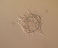 Amöbe mit einverleibtem Rädertierchen - Fokalebene 1 (8097059924).jpg 1,208 × 992; 330 KB
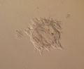 Amöbe mit einverleibtem Rädertierchen - Fokalebene 2 (8097061358).jpg 1,212 × 992; 400 KB
 Amöbe mit einverleibtem Rädertierchen - Fokalebene 3 (8097054847).jpg 1,208 × 996; 380 KB
 Amöbe? 400x, 12my - Fokalebene 1 (9394250407).jpg 931 × 913; 116 KB
 Amöbe? 400x, 12my - Fokalebene 1 (9394251411).jpg 931 × 913; 105 KB
 Amöbe? 400x, 12my - Fokalebene 1 (9394252413).jpg 931 × 913; 103 KB
 Amöbe? 400x, 12my - Fokalebene 1 (9397020810).jpg 931 × 913; 102 KB
 Amöbe? 400x, 12my - Gestackt (9394249365).jpg 931 × 913; 146 KB
 Biogen-components hg.jpg 2,587 × 2,070; 617 KB
 Dead Earwig with fungus.jpg 4,608 × 3,072; 2.78 MB
 Distribution of three-dimensional phototaxis in the tree of eukaryotes.jpg 1,280 × 784; 108 KB
 Divergence from a common ancestor.png 360 × 320; 91 KB
 Diversity of biomineralization across the eukaryotes.jpg 856 × 434; 247 KB
 Diversity of phototactic eukaryotes.jpg 1,639 × 1,800; 328 KB
 Ejemplos de plantas criptogamas.pdf 1,239 × 1,752, 2 pages; 316 KB
 Endosimbiosis.jpg 1,500 × 1,124; 129 KB
 Eukarya endosymbiosis.svg 563 × 282; 31 KB
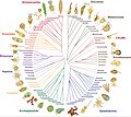 Eukaryota Arbol de vida.jpg 2,096 × 1,863; 854 KB
 Eukaryota diversity 2.jpg 804 × 919; 956 KB
 Eukaryote cell.jpg 2,743 × 1,576; 465 KB
 Eukaryote Phylogeny.png 1,569 × 876; 378 KB
 Eukaryote Tree of Life 2020.svg 607 × 582; 464 KB
 Eukaryotic tree of life (Burki et al 2020).jpg 3,381 × 1,724; 588 KB
 Foraminifera life cycle.png 2,826 × 1,580; 539 KB
 Goniomonas sp. - 1000x (10327848655).jpg 942 × 854; 398 KB
 Goniomonas sp. - 1000x (10327868925).jpg 968 × 874; 424 KB
 Il fungo Inotodus hispidus cresciuto su un albero di melo.jpg 4,032 × 3,024; 2.62 MB
 Infobox-Eukaryote.png 300 × 300; 87 KB
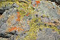 Lichen (7).jpg 4,608 × 3,072; 7.67 MB
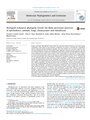 Molecular Phylogenetics and Evolution vol81 pp71-85.pdf 1,239 × 1,652, 15 pages; 2.77 MB
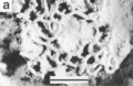 Occultammina sp.png 788 × 514; 434 KB
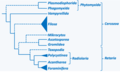 Phylogénie des Rhizaria - P.Silar 2016.png 879 × 527; 73 KB
 Picea abies crosses Haapastensyrjä Finland 2021-05-20.jpg 1,280 × 720; 319 KB
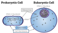 Prokaryotic vs eukaryotic cell.png 1,298 × 741; 342 KB
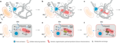 Proposed hypothetical model for eukaryogenesis.webp 2,153 × 769; 161 KB
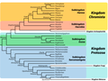 Protista taxonomy vs phylogeny.png 3,000 × 2,260; 512 KB
 Protists (10.3897-subtbiol.42.78037) Figure 4.jpg 1,512 × 1,249; 967 KB
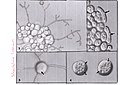 Schizochytrium limacinum..jpg 3,509 × 2,481; 3.17 MB
.jpg)


.jpg)
.jpg)
.jpg)
.jpg)
.jpg)
.jpg)
.jpg)
.jpg)


.jpg)

.jpg)
.jpg)


.jpg)


_Figure_4.jpg)
