Category:Flow cytometry
Zur Navigation springen
Zur Suche springen
Ein Messverfahren zur Analyse der Zellen in der Medizin und Biologie. | |||||
| Medium hochladen | |||||
| Unterklasse von |
| ||||
|---|---|---|---|---|---|
| |||||
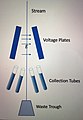
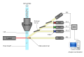
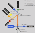
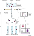
English: Flow cytometry is a method in cell biology that employs the deflection of laser light a well as the excitation of fluorescent dyes to analyse various properties of a high number cells in a relatively short time. This category lists methods and tools used in flow cytometry.
Unterkategorien
Es werden 4 von insgesamt 4 Unterkategorien in dieser Kategorie angezeigt:
In Klammern die Anzahl der enthaltenen Kategorien (K), Seiten (S), Dateien (D)
C
- Flow cytometers (5 D)
I
- Immunophenotyping (4 D)
Seiten in der Kategorie „Flow cytometry“
Diese Kategorie enthält nur die folgende Seite.
Medien in der Kategorie „Flow cytometry“
Folgende 200 Dateien sind in dieser Kategorie, von 340 insgesamt.
(vorherige Seite) (nächste Seite)-
-
-
-
-
-
-
-
-
A-Novel-Model-of-Intravital-Platelet-Imaging-Using-CD41-ZsGreen1-Transgenic-Rats-pone.0154661.s002.ogv 25 s, 1.024 × 348; 3,32 MB
-
A-Novel-Model-of-Intravital-Platelet-Imaging-Using-CD41-ZsGreen1-Transgenic-Rats-pone.0154661.s003.ogv 25 s, 1.024 × 348; 359 KB
-
-
-
-
-
-
-
A-Plasmodium-falciparum-Strain-Expressing-GFP-throughout-the-Parasite's-Life-Cycle-pone.0009156.s001.ogv 12 s, 1.388 × 1.040; 64 KB
-
A-Plasmodium-falciparum-Strain-Expressing-GFP-throughout-the-Parasite's-Life-Cycle-pone.0009156.s002.ogv 24 s, 1.388 × 1.040; 666 KB
-
-
-
-
An-intraductal-human-in-mouse-transplantation-model-mimics-the-subtypes-of-ductal-carcinoma-in-situ-bcr2358-S1.ogv 5 min 44 s, 320 × 240; 9,76 MB
-
Analysis-of-Cells-Targeted-by-Salmonella-Type-III-Secretion-In-Vivo-ppat.0030196.sv001.ogv 16 s, 558 × 524; 264 KB
-
Analysis-of-Cells-Targeted-by-Salmonella-Type-III-Secretion-In-Vivo-ppat.0030196.sv002.ogv 13 s, 542 × 476; 254 KB
-
Analysis-of-Cells-Targeted-by-Salmonella-Type-III-Secretion-In-Vivo-ppat.0030196.sv003.ogv 13 s, 542 × 476; 175 KB
-
Analysis-of-Cells-Targeted-by-Salmonella-Type-III-Secretion-In-Vivo-ppat.0030196.sv004.ogv 13 s, 542 × 476; 195 KB
-
Analysis-of-Cells-Targeted-by-Salmonella-Type-III-Secretion-In-Vivo-ppat.0030196.sv005.ogv 13 s, 542 × 476; 204 KB
-
-
-
-
-
-
 AutomatedFlowPipeline.svg 612 × 459; 238 KB
-
-
-
Biofilm-Induced-Tolerance-towards-Antimicrobial-Peptides-pone.0001891.s001.ogv 0,0 s, 512 × 480; 18,13 MB
-
-
-
-
-
 Cell Sorting Diagram.jpg 2.784 × 4.032; 10,04 MB
-
Cells-Expressing-the-CEBPbeta-Isoform-LIP-Engulf-Their-Neighbors-pone.0041807.s006.ogv 23 s, 640 × 480; 655 KB
-
Cells-Expressing-the-CEBPbeta-Isoform-LIP-Engulf-Their-Neighbors-pone.0041807.s007.ogv 53 s, 512 × 500; 1,99 MB
-
Cells-Expressing-the-CEBPbeta-Isoform-LIP-Engulf-Their-Neighbors-pone.0041807.s008.ogv 8,5 s, 1.392 × 1.040; 423 KB
-
Cells-Expressing-the-CEBPbeta-Isoform-LIP-Engulf-Their-Neighbors-pone.0041807.s009.ogv 13 s, 434 × 348; 131 KB
-
Cells-Expressing-the-CEBPbeta-Isoform-LIP-Engulf-Their-Neighbors-pone.0041807.s010.ogv 21 s, 530 × 512; 754 KB
-
Cells-Expressing-the-CEBPbeta-Isoform-LIP-Engulf-Their-Neighbors-pone.0041807.s011.ogv 25 s, 512 × 512; 276 KB
-
-
-
-
-
-
-
-
Circadian-Clocks-in-Mouse-and-Human-CD4+-T-Cells-pone.0029801.s002.ogv 12 s, 256 × 256; 2,15 MB
-
Contribution-of-Caspase(s)-to-the-Cell-Cycle-Regulation-at-Mitotic-Phase-pone.0018449.s002.ogv 24 s, 450 × 376; 318 KB
-
Contribution-of-Caspase(s)-to-the-Cell-Cycle-Regulation-at-Mitotic-Phase-pone.0018449.s003.ogv 24 s, 450 × 376; 426 KB
-
Contribution-of-Caspase(s)-to-the-Cell-Cycle-Regulation-at-Mitotic-Phase-pone.0018449.s004.ogv 24 s, 450 × 376; 470 KB
-
Contribution-of-Caspase(s)-to-the-Cell-Cycle-Regulation-at-Mitotic-Phase-pone.0018449.s005.ogv 24 s, 450 × 376; 428 KB
-
Contribution-of-Caspase(s)-to-the-Cell-Cycle-Regulation-at-Mitotic-Phase-pone.0018449.s006.ogv 24 s, 450 × 376; 477 KB
-
Contribution-of-Caspase(s)-to-the-Cell-Cycle-Regulation-at-Mitotic-Phase-pone.0018449.s007.ogv 49 s, 680 × 512; 2,09 MB
-
Contribution-of-Caspase(s)-to-the-Cell-Cycle-Regulation-at-Mitotic-Phase-pone.0018449.s008.ogv 49 s, 680 × 512; 1,41 MB
-
Crystallization-of-DNA-coated-colloids-ncomms8253-s2.ogv 21 s, 696 × 520; 26,32 MB
-
Crystallization-of-DNA-coated-colloids-ncomms8253-s3.ogv 23 s, 527 × 167; 776 KB
-
Crystallization-of-DNA-coated-colloids-ncomms8253-s4.ogv 18 s, 696 × 520; 15,03 MB
-
Crystallization-of-DNA-coated-colloids-ncomms8253-s5.ogv 15 s, 696 × 520; 20,47 MB
-
Crystallization-of-DNA-coated-colloids-ncomms8253-s6.ogv 17 s, 696 × 520; 23,61 MB
-
Crystallization-of-DNA-coated-colloids-ncomms8253-s7.ogv 17 s, 696 × 520; 27,44 MB
-
Crystallization-of-DNA-coated-colloids-ncomms8253-s8.ogv 20 s, 384 × 288; 11,91 MB
-
Crystallization-of-DNA-coated-colloids-ncomms8253-s9.ogv 20 s, 720 × 576; 14,23 MB
-
-
-
CXCR7-Functions-as-a-Scavenger-for-CXCL12-and-CXCL11-pone.0009175.s001.ogv 6,3 s, 335 × 311; 243 KB
-
CXCR7-Functions-as-a-Scavenger-for-CXCL12-and-CXCL11-pone.0009175.s002.ogv 5,7 s, 321 × 340; 186 KB
-
 Cytometer ru.svg 924 × 624; 77 KB
-
 Cytometer.svg 924 × 624; 65 KB
-
Cytotoxic-Effect-of-Poly-Dispersed-Single-Walled-Carbon-Nanotubes-on-Erythrocytes-In-Vitro-and-In-pone.0022032.s001.ogv 9,3 s, 1.280 × 1.024; 2,54 MB
-
-
-
-
-
-
Development-of-a-macromolecular-prodrug-for-the-treatment-of-inflammatory-arthritis-mechanisms-ar3130-S1.ogv 8,3 s, 992 × 1.040; 689 KB
-
-
Development-of-an-In-Vitro-Model-for-the-Multi-Parametric-Quantification-of-the-Cellular-pone.0032621.s011.ogv 1 min 10 s, 640 × 480; 11,91 MB
-
-
-
-
-
-
 Duchflusszytometer.png 3.160 × 3.035; 476 KB
-
-
-
-
-
-
-
Dynamics-of-Macrophage-Trogocytosis-of-Rituximab-Coated-B-Cells-pone.0014498.s003.ogv 4,0 s, 112 × 148; 48 KB
-
Dynamics-of-Macrophage-Trogocytosis-of-Rituximab-Coated-B-Cells-pone.0014498.s004.ogv 4,0 s, 224 × 572; 265 KB
-
Dynamics-of-Macrophage-Trogocytosis-of-Rituximab-Coated-B-Cells-pone.0014498.s005.ogv 3,9 s, 188 × 164; 233 KB
-
Dynamics-of-Macrophage-Trogocytosis-of-Rituximab-Coated-B-Cells-pone.0014498.s006.ogv 4,0 s, 352 × 208; 236 KB
-
Dynamics-of-Macrophage-Trogocytosis-of-Rituximab-Coated-B-Cells-pone.0014498.s007.ogv 4,0 s, 572 × 320; 336 KB
-
Dynamics-of-Macrophage-Trogocytosis-of-Rituximab-Coated-B-Cells-pone.0014498.s008.ogv 4,0 s, 200 × 500; 257 KB
-
Dynamics-of-Macrophage-Trogocytosis-of-Rituximab-Coated-B-Cells-pone.0014498.s009.ogv 0,7 s, 1.344 × 1.024; 871 KB
-
-
-
-
Entamoeba-histolytica-Cysteine-Proteinase-5-Evokes-Mucin-Exocytosis-from-Colonic-Goblet-Cells-via-ppat.1005579.s004.ogv 4,9 s, 1.104 × 1.104; 6,74 MB
-
Entamoeba-histolytica-Cysteine-Proteinase-5-Evokes-Mucin-Exocytosis-from-Colonic-Goblet-Cells-via-ppat.1005579.s005.ogv 4,5 s, 1.236 × 1.236; 4,43 MB
-
-
-
Extensive-Fusion-of-Mitochondria-in-Spinal-Cord-Motor-Neurons-pone.0038435.s001.ogv 5,8 s, 1.441 × 404; 570 KB
-
Extensive-Fusion-of-Mitochondria-in-Spinal-Cord-Motor-Neurons-pone.0038435.s002.ogv 6,8 s, 982 × 287; 209 KB
-
Extensive-Fusion-of-Mitochondria-in-Spinal-Cord-Motor-Neurons-pone.0038435.s003.ogv 5,9 s, 961 × 207; 134 KB
-
Extensive-Fusion-of-Mitochondria-in-Spinal-Cord-Motor-Neurons-pone.0038435.s004.ogv 18 s, 1.396 × 336; 678 KB
-
Extensive-Fusion-of-Mitochondria-in-Spinal-Cord-Motor-Neurons-pone.0038435.s005.ogv 18 s, 1.396 × 336; 923 KB
-
Extensive-Fusion-of-Mitochondria-in-Spinal-Cord-Motor-Neurons-pone.0038435.s006.ogv 18 s, 1.396 × 336; 1.021 KB
-
-
Externalized-decondensed-neutrophil-chromatin-occludes-pancreatic-ducts-and-drivespancreatitis-ncomms10973-s3.ogv 6,1 s, 1.388 × 1.040; 6,35 MB
-
-
-
Externalized-decondensed-neutrophil-chromatin-occludes-pancreatic-ducts-and-drivespancreatitis-ncomms10973-s6.ogv 7,6 s, 1.920 × 1.080; 3,53 MB
-
-
-
-
 Flow Cy.gif 336 × 322; 3 KB
-
 Flow cytometer structure.png 865 × 599; 1,98 MB
-
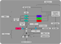 Flow cytometer.png 852 × 615; 2 MB
-
 Flow cytometric detection of plasma cells.png 512 × 108; 32 KB
-
 Flow cytometric gating by side scatter and CD45, without labels.png 1.451 × 839; 365 KB
-
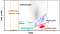 Flow cytometric gating by side scatter and CD45.png 1.459 × 837; 389 KB
-
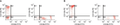
 Flow cytometric viability by 7-AAD.png 1.047 × 463; 73 KB
-
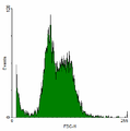
 Flow cytometry 3Dhistogram.png 375 × 256; 99 KB
-
 Flow cytometry histogram.png 266 × 268; 4 KB
-
 Flow cytometry scatterplot.png 285 × 288; 5 KB
-
 Flow cytometry scheme.png 592 × 766; 54 KB
-
Flow-Based-Cytometric-Analysis-of-Cell-Cycle-via-Simulated-Cell-Populations-pcbi.1000741.s007.ogv 50 s, 768 × 576; 2,67 MB
-
 FlowCAP Results.png 2.000 × 2.000; 361 KB
-
 FlowCitometer.svg 607 × 380; 19 KB
-
 Flowcytometer MoFlo (DAKO cytomation).jpg 320 × 426; 21 KB
-
 FlowCytometrySFLvsSSC.png 300 × 316; 35 KB
-
 Fluorescence Activated Cell Sorting (FACS) principle.tif 1.201 × 1.280; 380 KB
-
 Focalizzazione idrodinamica.png 883 × 750; 21 KB
-
Functional-classification-of-memory-CD8+-T-cells-by-CX3CR1-expression-ncomms9306-s8.ogv 12 s, 640 × 640; 568 KB
-
-
-
-
-
Genetic-epigenesis-pbio.1001325.s014.ogv 3,0 s, 1.548 × 616; 254 KB
-
Genetic-epigenesis-pbio.1001325.s015.ogv 3,0 s, 608 × 302; 48 KB
-
Genetic-epigenesis-pbio.1001325.s016.ogv 4,0 s, 400 × 540; 69 KB
-
Genetic-epigenesis-pbio.1001325.s017.ogv 7,3 s, 1.302 × 626; 187 KB
-
Genetic-epigenesis-pbio.1001325.s018.ogv 7,3 s, 1.302 × 526; 143 KB
-
High-density-lipoprotein-contribute-to-G0-G1S-transition-in-Swiss-NIH3T3-fibroblasts-srep17812-s1.ogv 4,0 s, 236 × 159; 1,13 MB
-
-
-
 HIV-AIDS-Immunstatusdiagnostik Kenia.JPG 4.912 × 3.264; 8,75 MB
-
-
IFlow-A-Graphical-User-Interface-for-Flow-Cytometry-Tools-in-Bioconductor-103839.f1.ogv 3 min 16 s, 808 × 624; 4,18 MB
-
-
-
In-Vitro-Formation-of--Cell-Pseudoislets-Using-Islet-Derived-Endothelial-Cells-pone.0072260.s001.ogv 1,4 s, 568 × 576; 364 KB
-
-
-
-
-
-
-
-
-
-
-
-
-
-
-
-
 Inside BD LSRFortessa cell analyzer.jpg 4.800 × 3.200; 701 KB
-
-
Lactobacillus-Decelerates-Cervical-Epithelial-Cell-Cycle-Progression-pone.0063592.s003.ogv 19 s, 694 × 520; 3,96 MB
-
Lactobacillus-Decelerates-Cervical-Epithelial-Cell-Cycle-Progression-pone.0063592.s004.ogv 19 s, 694 × 520; 7,11 MB
-
Lactobacillus-Decelerates-Cervical-Epithelial-Cell-Cycle-Progression-pone.0063592.s005.ogv 19 s, 694 × 520; 8,43 MB
-
Lactobacillus-Decelerates-Cervical-Epithelial-Cell-Cycle-Progression-pone.0063592.s006.ogv 19 s, 694 × 520; 3,83 MB
-
Large-scale-cell-production-of-stem-cells-for-clinical-application-using-the-automated-cell-1472-6750-13-102-S4.ogv 3 min 21 s, 400 × 300; 15,43 MB
-
-
-
-
-
-
-
Lineage-Tracking-for-Probing-Heritable-Phenotypes-at-Single-Cell-Resolution-pone.0152395.s008.ogv 30 s, 750 × 393; 2,72 MB
-
-
Long-term-maintenance-of-human-induced-pluripotent-stem-cells-by-automated-cell-culture-system-srep16647-s2.ogv 3 min 31 s, 426 × 240; 4,04 MB
-
-
-
-
-
-
-


.jpg)


