Category:Hippocampus (anatomy)
Jump to navigation
Jump to search
brain region correlated with memory consolidation and imagination | |||||
| Upload media | |||||
| Instance of |
| ||||
|---|---|---|---|---|---|
| Subclass of |
| ||||
| Part of | |||||
| |||||
The hippocampus is an anatomical subdivision of the brain. Hippocampus may also refer to:
- the seahorse genus. -> Category:Hippocampus
- Hippocamp, a mythological sea-horse. -> Category:Hippocampus (mythology)
Subcategories
This category has the following 13 subcategories, out of 13 total.
C
- Cornu Ammonis (9 F)
D
- Dentate gyrus (36 F)
F
- Fimbria of hippocampus (10 F)
G
- Grid cells (21 F)
H
- Hippocampal neurons (14 F)
- Hippocampal sulcus (9 F)
P
R
S
- Subgranular zone (4 F)
V
Pages in category "Hippocampus (anatomy)"
This category contains only the following page.
Media in category "Hippocampus (anatomy)"
The following 97 files are in this category, out of 97 total.
-
202102 Frontal plane of the brain hippocampus.svg 512 × 512; 259 KB
-
Amygdala hippocampus.jpg 769 × 564; 27 KB
-
Andersenův okruh.jpg 819 × 500; 46 KB
-
Brain cut section 2.jpg 960 × 540; 83 KB
-
Brain regions in memory formation updated.jpg 1,284 × 889; 122 KB
-
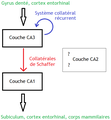
Connectivité axonale dans l'hippocampe.png 612 × 663; 20 KB
-
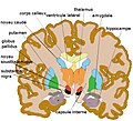
Cp coronale ssthal3.jpg 589 × 534; 74 KB
-
Depresión sináptica a largo plazo en el hipocampo.JPG 640 × 413; 34 KB
-
Depresión sináptica prolongada en el cerebelo.JPG 640 × 400; 42 KB
-
DGC dispersion.jpg 1,000 × 413; 128 KB
-
Diagram of a Timm-stained cross-section of the hippocampus.JPEG 1,542 × 842; 576 KB
-
Dopamine and serotonin pathways ar.png 720 × 480; 86 KB
-
Dopamine and serotonin pathways-es.png 720 × 480; 79 KB
-
Dopamine and serotonin pathways.png 720 × 480; 55 KB
-
Dopamine pathways -ru.svg 479 × 335; 27 KB
-
Dopamine pathways ar.svg 479 × 335; 88 KB
-
Dopamine Pathways vie.png 450 × 334; 104 KB
-
Dopamine pathways zh-hans.svg 479 × 335; 27 KB
-
Dopamine pathways zh-hant.svg 479 × 335; 27 KB
-
Dopamine Pathways-es.png 500 × 371; 148 KB
-
Dopamine Pathways.png 450 × 334; 148 KB
-
Dopamine pathways.svg 479 × 335; 27 KB
-
Dopamine serotonin rus.png 1,665 × 1,014; 463 KB
-
Epilepsy- right hippocampal seizure onset.png 435 × 226; 71 KB
-
Formación hipocampal2-vi.svg 102 × 122; 46 KB
-
Formación hipocampal2.svg 102 × 122; 126 KB
-
Gehirn Frontalschnitt hippocampus-it.png 1,055 × 573; 247 KB
-
Gehirn Frontalschnitt hippocampus.png 913 × 573; 263 KB
-
GPS-in-the-brain.png 5,562 × 2,922; 22.99 MB
-
Gray717.png 500 × 568; 102 KB
-
Gray739-emphasizing-calcar-avis.png 1,000 × 758; 997 KB
-
Gray739-emphasizing-hippocampus-IT.png 500 × 379; 240 KB
-
Gray739-emphasizing-hippocampus-minor.png 1,000 × 758; 997 KB
-
Gray739-emphasizing-hippocampus-vi.png 500 × 420; 241 KB
-
Gray739-emphasizing-hippocampus.png 500 × 379; 263 KB
-
Gray739.png 500 × 379; 186 KB
-
Gray740.png 266 × 494; 52 KB
-
Gray747.png 450 × 310; 17 KB
-
Gray748.png 500 × 510; 66 KB
-
Gray749.png 500 × 308; 22 KB
-
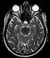
Hippocampal sulcus remnants - MRT T2 axial - 001.jpg 1,437 × 1,663; 192 KB
-
Hippocampale Sulcusreste rechts 68W - MR T2 axial - 001 - Annotation.jpg 1,440 × 1,631; 160 KB
-
Hippocampale Sulcusreste rechts 68W - MR T2 axial - 001.jpg 1,440 × 1,631; 237 KB
-
Hippocampe parahippo.png 1,000 × 800; 426 KB
-
Hippocampus (Brain) Unlabeled.jpg 1,024 × 881; 195 KB
-
Hippocampus (brain).jpg 1,024 × 881; 62 KB
-
Hippocampus - DK ATLAS.png 1,200 × 900; 490 KB
-
Hippocampus and seahorse cropped.JPG 798 × 511; 72 KB
-
Hippocampus and seahorse.JPG 2,274 × 1,700; 162 KB
-
Hippocampus coronal section176157.fig.004-vi.jpg 600 × 531; 126 KB
-
Hippocampus coronal section176157.fig.004.jpg 600 × 531; 152 KB
-
Hippocampus coronal sections.gif 148 × 158; 1.18 MB
-
Hippocampus image.png 800 × 455; 310 KB
-
Hippocampus Layering Wiring.tif 2,261 × 3,183; 2.63 MB
-
Hippocampus macroscopy.jpg 448 × 338; 31 KB
-
Hippocampus sagittal sections.gif 185 × 158; 1.21 MB
-
Hippocampus scheme.jpg 1,128 × 658; 161 KB
-
Hippocampus small.gif 200 × 200; 563 KB
-
Hippocampus transversal sections.gif 148 × 185; 1.22 MB
-
Hippocampus weights.png 832 × 1,093; 1.15 MB
-
Hippocampus-Bird brain.png 2,800 × 2,066; 2.2 MB
-
Hippocampus-layout-schema.png 801 × 1,029; 155 KB
-
Hippocampus-mri.jpg 510 × 510; 71 KB
-
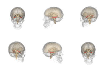
Hippocampus.gif 600 × 600; 4.09 MB
-
Hippocampus.png 231 × 274; 39 KB
-
Hippocampus.svg 254 × 295; 258 KB
-
Hippokampus.jpg 720 × 540; 53 KB
-
Hippolobes it.gif 400 × 261; 55 KB
-
Hippsysteem.PNG 582 × 534; 16 KB
-
Hmstradecki hippocampus migration.png 916 × 625; 70 KB
-
Human brain right dissected lateral view description.JPG 653 × 413; 40 KB
-
Human hippocampus.png 320 × 248; 43 KB
-
Human temporal lobe areas.png 1,793 × 1,513; 1.63 MB
-
Le doux.png 569 × 545; 13 KB
-
Left Hippocampal Sclerosis on MRI.jpg 1,280 × 687; 131 KB
-
Major gray and white matter limbic structures.jpg 850 × 592; 63 KB
-
-
-
Neural systems proposed to process emotion.png 945 × 461; 314 KB
-
Neuronal connections of nucleus accumbens.JPEG 1,093 × 1,093; 78 KB
-
Paden.PNG 748 × 402; 23 KB
-
Parahippocampal gyrus - inferiror view.png 536 × 638; 280 KB
-
Place cel remapping.png 745 × 561; 70 KB
-
S-ART Mindfulness and brain1.jpg 843 × 585; 369 KB
-
Sobo 1909 637.png 1,061 × 1,050; 3.19 MB
-
Sobo 1909 639.png 564 × 806; 1.74 MB
-
Sobo 1909 640.png 739 × 488; 1.03 MB
-
Sobo 1909 641.png 1,027 × 730; 2.15 MB
-
Sobo 1909 646.png 1,201 × 773; 2.66 MB
-
Subcortical structures.png 646 × 200; 129 KB
-
Vestibular cortices and spatial cognition.jpg 569 × 850; 333 KB
-
-
WholeCellPatchClamp-03.jpg 1,344 × 1,024; 345 KB
-
WholeCellPatchClamp-03inv.jpg 1,344 × 1,024; 465 KB


