Category:Histology
Mine navigeerimisribale
Mine otsikasti

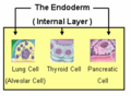
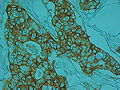
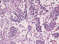
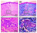
Histology is the study of the microscopic anatomy of cells and tissues of plants and animals. Place files relating to normal, disease-free tissues in a suitable subcategory of "Category:Animal histology", "Category:Human histology" or "Category:Plant histology".
study of the microscopic anatomy of cells and tissues of plants and animals  | |||||
| Laadi fail üles | |||||
| Üksikjuht nähtusest | |||||
|---|---|---|---|---|---|
| Mille alamklass | |||||
| Mitte segamini ajada | |||||
| |||||
- Place files relating to diseased tissues in a suitable subcategory of "Category:Histopathology":
- Diseased tissues of human beings should be placed in a suitable subcategory of "Category:Human histopathology".
- Diseased tissues of animals should be placed in a suitable subcategory of "Category:Veterinary histopathology".
Alamkategooriad
Järgmised 31 alamkategooriat on selles kategoorias (kokku 31).
*
.
- BrainMaps (10 P)
?
A
B
C
D
- Dye carrier (8 P)
F
- Fixation (histology) (2 P)
G
H
- Histology of apoptosis (2 P)
I
M
P
- Histology of peritoneum (2 P)
S
T
Failid kategoorias "Histology"
Järgmised 200 faili on selles kategoorias (kokku 367).
(eelmine lehekülg) (järgmine lehekülg)-
 20090903-153949a (3891900480).jpg 720 × 480; 127 KB
-
 20180427SpinalCord11stack (26875138687).jpg 3360 × 2832; 5,76 MB
-
 201904 myotube.svg 512 × 410; 182 KB
-
 A thinking neurocomputer.jpg 1069 × 1313; 568 KB
-
 Actinomices (citología de cuello uterino) (9439770787).jpg 1280 × 960; 483 KB
-
 Actinomices (citología de cuello uterino) (9439770817).jpg 1280 × 960; 428 KB
-
 Actinomices (citología de cuello uterino) (9439771045).jpg 1280 × 960; 353 KB
-
 Actinomices (citología de cuello uterino) (9439771101).jpg 1280 × 960; 367 KB
-
 Actinomices (citología de cuello uterino) (9442555742).jpg 1280 × 960; 441 KB
-
 Actinomices (citología de cuello uterino) (9442555744).jpg 1280 × 960; 456 KB
-
 Actinomices (citología de cuello uterino) (9442555752).jpg 1280 × 960; 411 KB
-
 Actinomices (citología de cuello uterino) (9442556076).jpg 1280 × 960; 367 KB
-
 Actinomycosis histology.jpg 2336 × 2829; 2,45 MB
-
 Actinomycosis.pdf 1239 × 1752; 988 KB
-
 Adenoma.jpg 2073 × 1046; 350 KB
-
 Amyloidosis1.gif 247 × 187; 43 KB
-
 An almost intelligent ocean.jpg 914 × 1338; 616 KB
-
 Anexin stain (9734833573).jpg 1347 × 1013; 330 KB
-
 Angiomatosis hemangiomatosis Case 277 (9723365427).jpg 2272 × 1704; 1,47 MB
-
 Apparatus for preparing injected preparations Wellcome M0018212.jpg 2717 × 3889; 1,59 MB
-
 ArteryVeinCS.jpg 640 × 480; 187 KB
-
 ASCH, con vaginosis (9392539118).jpg 1280 × 960; 432 KB
-
 ASCH, con vaginosis (9392539154).jpg 1280 × 960; 487 KB
-
 ASCH, con vaginosis (9392539160).jpg 1280 × 960; 383 KB
-
 Atipia de células escamosas (ASC, ASCUS) (9292924153).jpg 1280 × 960; 322 KB
-
 Atipia de células escamosas (ASCUS) (9412585837).jpg 1280 × 960; 418 KB
-
 Atipia de células escamosas (ASCUS) (9415351776).jpg 1280 × 960; 397 KB
-
 Atipia de células escamosas (ASCUS, ASC) (9392112063).jpg 1280 × 960; 419 KB
-
 Atipia de células escamosas (ASCUS, ASC) (9392112125).jpg 1280 × 960; 311 KB
-
 Atipia de células escamosas (ASCUS, ASC) (9392112175).jpg 1280 × 960; 365 KB
-
 Atipia de células escamosas (ASCUS, ASC) (9392112465).jpg 1280 × 960; 426 KB
-
 Atipia de células escamosas (ASCUS, ASC) (9392112495).jpg 1280 × 960; 411 KB
-
 Atipia de células escamosas (ASCUS, ASC) (9392112725).jpg 1280 × 960; 447 KB
-
 Atipia de células escamosas (ASCUS, ASC) (9392112727).jpg 1280 × 960; 425 KB
-
 Atipia de células escamosas (ASCUS, ASC) (9394881622).jpg 1280 × 960; 432 KB
-
 Atipia de células escamosas (ASCUS, ASC) (9394881924).jpg 1280 × 960; 467 KB
-
 Atipia de células escamosas (ASCUS, ASC) (9394882140).jpg 1280 × 960; 418 KB
-
 Atipia de células escamosas no descartable HSIL (ASC-H) (9439523243).jpg 1280 × 960; 335 KB
-
 Atipia de células escamosas no descartable HSIL (ASC-H) (9442307718).jpg 1280 × 960; 365 KB
-
 Atipia de células escamosas, probable LSIL, con vaginosis. (9130291917).jpg 1280 × 960; 335 KB
-
 Atipia de células escamosas, probable LSIL, con vaginosis. (9132501506).jpg 1280 × 960; 425 KB
-
 Atipia de ´células escamosas (ASC, ASCUS) (9292923841).jpg 1280 × 960; 329 KB
-
 Atipia de ´células escamosas (ASC, ASCUS) (9292923849).jpg 1280 × 960; 331 KB
-
 Atipia de ´células escamosas (ASC, ASCUS) (9292923873).jpg 1280 × 960; 314 KB
-
 Atipia de ´células escamosas (ASC, ASCUS) (9292924085).jpg 1280 × 960; 320 KB
-
 Atipia de ´células escamosas (ASC, ASCUS) (9295702360).jpg 1280 × 960; 315 KB
-
 Basal cell carcinoma histology image.jpg 2464 × 3188; 3,04 MB
-
 Blue Intelligent Ocean Yury Scherbatykh.jpg 1152 × 1718; 383 KB
-
 Blue leopards of the spinal cord.jpg 1113 × 1370; 753 KB
-
 Box of 126 microscope preparations of a spinal column, Edinb Wellcome L0057901.jpg 2832 × 4256; 1,04 MB
-
 Boîte de coupes histologiques (physiologie) - IHM-0651.jpg 3600 × 2612; 2,09 MB
-
 Boîte de coupes histologiques - IHM-0254.jpg 3088 × 2056; 2,9 MB
-
 Brockhaus and Efron Encyclopedic Dictionary b65 328-1.jpg 3135 × 2625; 1,52 MB
-
 Brockhaus and Efron Encyclopedic Dictionary b65 328-2.jpg 3403 × 2774; 1,72 MB
-
 Brockhaus and Efron Encyclopedic Dictionary b65 328-3.jpg 2953 × 2563; 1,01 MB
-
 Brockhaus and Efron Encyclopedic Dictionary b65 328-4.jpg 3554 × 2803; 924 KB
-
 Brockhaus and Efron Encyclopedic Dictionary b65 328-5.jpg 1594 × 2277; 317 KB
-
 Brown tumour - intermed mag.jpg 4272 × 2848; 4,81 MB
-
 Bulbe de Mastophora mâle.jpg 4027 × 2597; 848 KB
-
 Cambium2.jpg 480 × 324; 62 KB
-
 CardiacMuscle - longtitudinal.jpg 640 × 480; 188 KB
-
 Cartoon Hyaline Cartilage.jpg 2049 × 1600; 176 KB
-
 Cellules, tissus, organes et systèmes.jpg 1754 × 2480; 842 KB
-
 Cerebral cortex slide.jpg 588 × 529; 203 KB
-

-
 CHL lacunar cell x40.jpg 1040 × 772; 454 KB
-
 Chromo.jpg 561 × 384; 65 KB
-
 Chronic inflammation slide.jpg 540 × 568; 261 KB
-
 CIN 3, Liquid-based Pap (3995927839).jpg 1414 × 944; 534 KB
-
 Circumvallate.jpg 2048 × 1536; 595 KB
-
 Citoesqueleto.gif 355 × 256; 51 KB
-
 Cluster (rus).jpg 1296 × 433; 151 KB
-
 Cluster (ukr).jpg 2592 × 866; 399 KB
-
 Colangiocarcinoma intrahepático.jpg 2592 × 1944; 3,95 MB
-
 Comparison of cancer cell lines.png 640 × 285; 11 KB
-
 Conger type callus 3ms White Light.TIF 2048 × 1536; 9,01 MB
-
 Coupe totale d'araignée.jpg 1743 × 1122; 217 KB
-
 Cross section normal nerve and atrophied nerve.png 992 × 476; 633 KB
-
 Cryostat Stage.jpg 1632 × 1224; 720 KB
-
 Cryptosporidium parvum auramine-rhodamine labeled.jpg 300 × 308; 6 KB
-
 Cuboidal Epithelium Section.jpg 640 × 480; 338 KB
-

-
 Decalcified Compact Bone slide.jpg 2048 × 1536; 650 KB
-
 DenseConnectiveTissue-1.jpg 640 × 480; 184 KB
-

-
 Elastic CT H&E.jpg 1502 × 1476; 933 KB
-
 Elastic CT VVG.jpg 634 × 679; 228 KB
-
 Elastic-Orcein.jpg 2048 × 1536; 300 KB
-
 Elastic-VVG.jpg 2048 × 1536; 454 KB
-
 ElastinisJ.A.JPG 1332 × 1200; 393 KB
-
 Endocrinoide mâle 2.jpg 495 × 325; 32 KB
-
 Endoderm2 hr.png 400 × 290; 62 KB
-
 Endoderm2-ar.png 270 × 198; 30 KB
-
 Endoderm2.png 270 × 198; 37 KB
-
 Epidermis histology 2014.jpg 4912 × 3264; 710 KB
-
 ErbB2.jpg 2048 × 1536; 779 KB
-
 FatStemCells.gif 260 × 208; 29 KB
-
 Female urethra histology.jpg 4912 × 3264; 994 KB
-
 Figures showing cell division. Wellcome M0016974.jpg 5060 × 2156; 2,73 MB
-
 Filistata insidiatrix, région épigastrique.jpg 3581 × 2525; 766 KB
-
 Foreign body.jpg 2272 × 1704; 2,86 MB
-
 Freezing microtome, London, England, 1883-1885 Wellcome L0058209.jpg 4256 × 2832; 1,25 MB
-
 Frozen tissue array block.jpg 3072 × 2304; 1,04 MB
-
 Frozen tissue array section.jpg 1077 × 680; 252 KB
-
 Fundus part of stomach.jpg 2288 × 1065; 855 KB
-
 G cell miguelferig.png 1404 × 1393; 95 KB
-
 GAFA 1996-01-3-1 Str 51 Bone 200fach XPL.jpg 2592 × 1944; 1,15 MB
-
 Glande clypéale d' Argyrodes zonatus.jpg 1849 × 2794; 597 KB
-
 Glande rostrale de Diguetia.jpg 4171 × 3427; 892 KB
-
 Glande venin Diguetia 1.jpg 3525 × 2430; 1,1 MB
-
 Glande venin Diguetia 2.jpg 1836 × 1237; 363 KB
-
 Glande venin Diguetia 3.jpg 1867 × 1179; 405 KB
-
 Glandes Scytodes 1.jpg 4312 × 3417; 815 KB
-
 Glandes Scytodes 2.jpg 5029 × 3370; 1,07 MB
-
 Glandula sebasea (3) - copia.jpg 529 × 592; 203 KB
-
 Glandular epithelium of small intestine (crypts of Lieberkühn).jpg 2473 × 1944; 5,1 MB
-
 Glandular tissue (small intestine).jpg 1232 × 1232; 1,84 MB
-
 GlaudusisAudinys.JPG 1600 × 1200; 377 KB
-
 Glazed eyes.jpg 1120 × 1426; 697 KB
-
 Globet cell miguelferig.png 921 × 1395; 98 KB
-
 Globus pallidus and putamen - very low mag.jpg 4272 × 2848; 5,93 MB
-
 Glomerulus.jpg 1500 × 1500; 191 KB
-
 Glycogen slide.jpg 348 × 341; 100 KB
-
 Golgy complex slide1.jpg 406 × 395; 109 KB
-
 Golgy complex slide2.jpg 387 × 436; 100 KB
-
 Grasa parda vista al microscopio electrónico.tif 676 × 500; 1,11 MB
-
 Gray964-zh.png 402 × 600; 154 KB
-
 Haematoxylin powder.jpg 2716 × 2673; 1,28 MB
-
 HE cystic renal dysplasia.jpg 2448 × 1920; 2,63 MB
-
 HE Microgyria cerebellum.jpg 2448 × 1920; 2,34 MB
-
 HE myocardial infarct with neutrophils infiltration.jpg 2448 × 1920; 3,06 MB
-
 HE Neuroblastoma beginning of differentiation toward ganglion cells.jpg 2448 × 1920; 2,24 MB
-
 HE Neuroblastoma Homer-Wright rosettes.jpg 2448 × 1920; 2,28 MB
-
 HE wall of bronchogenic cyst.jpg 2448 × 1920; 1,74 MB
-
 Healthy mammary gland.jpg 250 × 186; 31 KB
-
 Hem.jpg 304 × 228; 42 KB
-
 Heme.jpg 304 × 228; 41 KB
-
 HepG2.jpg 2816 × 2112; 1,55 MB
-
 Hersilia sp., région épigastrique.jpg 3165 × 2074; 656 KB
-
 Hiperplasia wiki.PNG 644 × 234; 358 KB
-
 Hipotalamo Parvo Magnocelular.png 744 × 636; 925 KB
-
 Histol.Technik.jpg 3636 × 1364; 1,01 MB
-
 Histologi Hepar.jpg 1024 × 1024; 108 KB
-
 Histologi kelenjar.jpg 1500 × 1500; 203 KB
-
 Histologi Organ.jpg 1024 × 1024; 77 KB
-
 Histologi Otot.jpg 1600 × 1600; 237 KB
-
 Histologi paru-paru.jpg 1024 × 768; 95 KB
-
 Histologi sistem pencernaan.jpg 1024 × 1024; 144 KB
-
 Histologi Tulang.jpg 1024 × 1024; 76 KB
-
 Histologi.jpg 1600 × 1600; 340 KB
-
 Histologia.jpg 977 × 1239; 773 KB
-
 Histologic Slide under MIcroscope.jpg 2848 × 4288; 2,84 MB
-
 Histological Architecture of the Joint Surface.jpg 576 × 359; 34 KB
-
 Histologie (1).jpg 2140 × 1027; 1,01 MB
-
 Histologie (3).jpg 4937 × 2250; 1,51 MB
-
 Histologie Basalmembran (2).jpg 2149 × 1226; 679 KB
-
 Histologie Basalmembran.jpg 2149 × 1226; 674 KB
-
 Histology (1).jpg 2700 × 1800; 966 KB
-
 Histology image of testis.jpg 1800 × 4000; 1,68 MB
-
 Histology Lab.jpg 5616 × 3744; 432 KB
-
 Histology of Adipose tissue.jpg 2472 × 1445; 1,33 MB
-
 Histology of cardiac muscle.jpg 2403 × 1467; 1,35 MB
-
 Histology of Ileum.png 1200 × 1600; 1,91 MB
-
 Histology of jejunum.jpg 3000 × 2244; 3,19 MB
-
 Histology of keratinised epithelium.jpg 2863 × 1589; 1,73 MB
-
 Histology of mixed salivary gland.jpg 1200 × 1600; 124 KB
-
 Histology of pseudo stratified ciliates columnar epithelium.jpg 2326 × 1367; 1,13 MB
-
 Histology of pulp calcifications.jpg 2807 × 1700; 777 KB
-
 Histology of simple columnar epithelium.jpg 3032 × 1886; 1,99 MB
-
 Histology of smooth muscle.jpg 2488 × 1447; 1,11 MB
-
 Histology of stratified squamous non keratinised epithelium.jpg 2283 × 1365; 1,17 MB
-
 Histology of transitional epithelium.jpg 2577 × 1402; 1,32 MB
-
 Histology RGNT HE.jpg 2080 × 1542; 796 KB
-
 Histology smooth muscle.jpg 2879 × 1640; 1,43 MB
-
 Histology tissue culture (1).jpg 2701 × 1600; 1,19 MB
-
 Histology tissue culture.jpg 1644 × 2701; 1,08 MB
-
 Histology.jpg 2700 × 1800; 1,19 MB
-
 Hydracs100x.jpg 1024 × 768; 170 KB
-
 Hydracs40x.jpg 1024 × 768; 134 KB
-
 Hypersegmented PMN.JPG 2272 × 1704; 1,11 MB
-
 Illu testis schematic.jpg 212 × 250; 37 KB
-
 Incomplete fixation.jpg 1969 × 2689; 1,11 MB
-
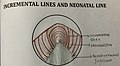 Incremental lines and neonatal line.jpg 2668 × 1462; 1,52 MB
-
 Induction-of-neocollagenesis-with-ellanse.jpg 726 × 647; 231 KB
-
 Injected kidney Gelatin carmine.jpg 476 × 490; 100 KB
-
 Intervertebral disks.jpg 960 × 720; 142 KB
-
 Jejunum.png 1200 × 1600; 1,98 MB
-
 Jonction serr.png 513 × 473; 30 KB
-
 Keloid slide.jpg 540 × 572; 169 KB
-
 Keratin.jpg 180 × 166; 9 KB
-
 Kidney Glomerulus Cell Types.png 989 × 1125; 1,17 MB
-
 Kidney H&E.jpg 453 × 503; 118 KB
-
 King's College of Household and Social Science 1930.png 541 × 670; 442 KB
-
 Kompakt (Kortikal) Kemik Histolojisi.jpg 921 × 1322; 1,35 MB
-
 Langerhans cell p.gif 350 × 335; 12 KB
-
 Large intestine histology.jpg 840 × 1271; 801 KB
-
 Large vein.jpg 1216 × 912; 100 KB
-
 LDOC1L Stain.png 599 × 599; 767 KB
-
 Le Gros Clarks CNS demonstration slides box.jpg 3600 × 2855; 3,27 MB
-
 Leberazinus.jpg 9836 × 3903; 12,5 MB
.jpg)
.jpg)


_(9439770787).jpg)
_(9439770817).jpg)
_(9439771045).jpg)
_(9439771101).jpg)
_(9442555742).jpg)
_(9442555744).jpg)
_(9442555752).jpg)
_(9442556076).jpg)


.jpg)
.jpg)


.jpg)
.jpg)
.jpg)
_(9292924153).jpg)
_(9412585837).jpg)
_(9415351776).jpg)
_(9392112063).jpg)
_(9392112125).jpg)
_(9392112175).jpg)
_(9392112465).jpg)
_(9392112495).jpg)
_(9392112725).jpg)
_(9392112727).jpg)
_(9394881622).jpg)
_(9394881924).jpg)
_(9394882140).jpg)
_(9439523243).jpg)
_(9442307718).jpg)
.jpg)
.jpg)
_(9292923841).jpg)
_(9292923849).jpg)
_(9292923873).jpg)
_(9292924085).jpg)
_(9295702360).jpg)


_-_IHM-0651.jpg)


.jpg)


_(9454839401).jpg)


_-_copia.jpg)
.jpg)
.jpg)


.jpg)
.jpg)
.jpg)

.jpg)


.jpg)


_Kemik_Histolojisi.jpg)


.jpg)
.jpg)