Category:Immunohistochemistry
Jump to navigation
Jump to search
common application of immunostaining | |||||
| Upload media | |||||
| Instance of |
| ||||
|---|---|---|---|---|---|
| Subclass of |
| ||||
| Part of |
| ||||
| |||||
Subcategories
This category has the following 4 subcategories, out of 4 total.
C
I
- Immunohistochemical analysis (41 F)
Pages in category "Immunohistochemistry"
This category contains only the following page.
Media in category "Immunohistochemistry"
The following 100 files are in this category, out of 100 total.
-
-
-
-
-
-
-
Androgen receptors on a HGSC tumour.jpg 215 × 176; 12 KB
-
-
BrdU staining (green) of radial glia.jpg 1,200 × 608; 275 KB
-
Cell flower.jpg 1,573 × 1,181; 211 KB
-
Connexion of neuves.jpg 1,376 × 1,032; 126 KB
-
Cutaneous Leiomyoma (Desmin Immunostain) (4344444718).jpg 1,745 × 1,291; 930 KB
-
Desert Rose.png 2,048 × 2,048; 9.19 MB
-
-
-
-
-
-
-
-
-
E-Cadherin Immunostain, Control to Show Typical Membranous Staining (14177993315).jpg 1,539 × 1,354; 555 KB
-
Fetal-Myocardium-in-the-Kidney-Capsule-An-In-Vivo-Model-of-Repopulation-of-Myocytes-by-Bone-Marrow-pone.0031099.s001.ogv 13 s, 1,596 × 1,080; 14.08 MB
-
Flock of Birds.jpg 2,256 × 2,256; 2.29 MB
-
FTLD TSP43 hippocampus.jpg 2,080 × 1,542; 905 KB
-
Gluten digestion.PNG 524 × 358; 17 KB
-
Her2neu 3+staining.jpg 4,912 × 3,684; 3.68 MB
-
-
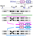
HLA-DQ locus.png 378 × 424; 18 KB
-
HLA-DQ protein.PNG 420 × 424; 15 KB
-
Hypothalamus of a mouse tissue stained by ABC-Immunohistochemistry.jpg 1,350 × 1,196; 947 KB
-
IHC CMV CMV lungs.jpg 2,448 × 1,920; 1.84 MB
-
IHC-1.jpg 371 × 498; 14 KB
-
IHC-FITC.jpg 567 × 646; 18 KB
-
Img398.C9 en infarto.jpg 1,604 × 1,009; 149 KB
-
Immunohistochemicalstaining1.PNG 368 × 176; 4 KB
-
Immunohistochemicalstaining2.PNG 368 × 211; 5 KB
-
Inmunofluorescencia-directa.svg 172 × 293; 99 KB
-
Intruders.jpg 1,376 × 1,032; 45 KB
-
Invasive Lobular Carcinoma of the Breast (13991322939).jpg 1,204 × 1,030; 318 KB
-
Islet-Formation-during-the-Neonatal-Development-in-Mice-pone.0007739.s005.ogv 26 s, 1,284 × 771; 4.66 MB
-
Larval White Seabass (3 days post hatching) .tif 6,000 × 4,000; 68.68 MB
-
LIN28 ependymoblastoma.jpg 2,080 × 1,542; 771 KB
-
Low-grade NET-008, synaptophysin.jpg 1,660 × 1,105; 552 KB
-
Luteoma of Pregnancy (inhibin immunostain) (4745655423).jpg 1,672 × 1,178; 862 KB
-
-
-
-
-
-
Mouse-Embryonic-Retina-Delivers-Information-Controlling-Cortical-Neurogenesis-pone.0015211.s002.ogv 33 s, 640 × 360; 1.53 MB
-
Neon cells.jpg 1,376 × 1,032; 591 KB
-
Neuron in the microchannel of hydrogel implant.png 5,240 × 1,034; 9.97 MB
-
-
Neuropathology case V 04.jpg 2,080 × 1,542; 822 KB
-
Neuropathology case V 05.jpg 2,080 × 1,542; 824 KB
-
Neuropathology case V 06.jpg 2,080 × 1,542; 826 KB
-
Neuropathology case VII 06.jpg 2,080 × 1,542; 685 KB
-
Neuropathology case VIII 04.jpg 2,080 × 1,542; 695 KB
-
Neuropathology case VIII 05.jpg 2,080 × 1,542; 749 KB
-
Neurothekeoma, S-100 immunostain (6813033020).jpg 1,470 × 1,469; 557 KB
-
Neurothekeoma, S-100 immunostain (6813033048).jpg 1,456 × 1,455; 526 KB
-
NP astrocyte GFAP.jpg 2,080 × 1,542; 790 KB
-
Purkinje C17orf75.png 624 × 660; 855 KB
-
PXA GFAP IHC.jpg 2,080 × 1,542; 760 KB
-
Retina and.jpg 1,304 × 977; 472 KB
-
RGNT Synapto.jpg 2,080 × 1,542; 960 KB
-
Schwannoma Histopathology S100.jpg 4,912 × 3,684; 5.08 MB
-
Schwannoma, S-100 Immunostain (5203888371).jpg 1,672 × 1,114; 739 KB
-
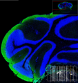
Section of mouse brain (false color).png 2,048 × 2,048; 8.53 MB
-
Self-Organizing-3D-Human-Neural-Tissue-Derived-from-Induced-Pluripotent-Stem-Cells-Recapitulate-pone.0161969.s005.ogv 4.5 s, 1,024 × 1,024; 3.77 MB
-
Self-Organizing-3D-Human-Neural-Tissue-Derived-from-Induced-Pluripotent-Stem-Cells-Recapitulate-pone.0161969.s006.ogv 3.7 s, 1,024 × 1,024; 1.32 MB
-
Small cell carcinoma, CAM 5.2 immunostain (1000x) (321570768).jpg 1,024 × 768; 325 KB
-
Small cell carcinoma, CAM 5.2 immunostain (400x) (321570771).jpg 1,024 × 768; 330 KB
-
Targeting-MT1-MMP-as-an-ImmunoPET-Based-Strategy-for-Imaging-Gliomas-pone.0158634.s004.ogv 9.7 s, 736 × 736; 3.19 MB
-
Targeting-MT1-MMP-as-an-ImmunoPET-Based-Strategy-for-Imaging-Gliomas-pone.0158634.s005.ogv 9.7 s, 560 × 560; 2.36 MB
-
Tela de araña.jpg 857 × 321; 272 KB
-
-
Tissue-and-Stage-Specific-Distribution-of-Wolbachia-in-Brugia-malayi-pntd.0001174.s002.ogv 8.2 s, 352 × 288; 62 KB
-
Tissue-and-Stage-Specific-Distribution-of-Wolbachia-in-Brugia-malayi-pntd.0001174.s003.ogv 8.2 s, 352 × 288; 79 KB
-
Tissue-and-Stage-Specific-Distribution-of-Wolbachia-in-Brugia-malayi-pntd.0001174.s004.ogv 8.2 s, 320 × 240; 54 KB
-
Uniqueness.png 2,048 × 2,048; 7.96 MB
-
-
-
-
-
-
-
Viral-Epidemics-in-a-Cell-Culture-Novel-High-Resolution-Data-and-Their-Interpretation-by-a-pone.0015571.s012.ogv 42 s, 1,360 × 1,024; 1,012 KB
-
-
Viral-Epidemics-in-a-Cell-Culture-Novel-High-Resolution-Data-and-Their-Interpretation-by-a-pone.0015571.s014.ogv 20 s, 1,250 × 1,082; 8.48 MB
-
Well differentiated NET bone metastasis Synaptophysin х400.jpg 4,032 × 3,024; 2.79 MB
-
Непрямой иммуногистохимический метод.jpg 368 × 211; 30 KB
-
Прямой иммуногистохимический метод.jpg 368 × 176; 24 KB


