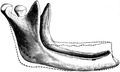
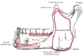
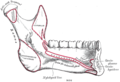
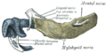
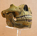
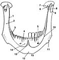
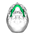
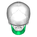
Category:Human mandible
Bước tới điều hướng
Bước tới tìm kiếm
 Human anatomy Mandible
| ||
|---|---|---|
lower jaw bone   | |||||
| Tải lên phương tiện | |||||
| Là một |
| ||||
|---|---|---|---|---|---|
| Là tập hợp con của |
| ||||
| Một phần của | |||||
| Nối liền | |||||
| Khác với | |||||
| |||||
Thể loại con
Thể loại này có 20 thể loại con sau, trên tổng số 20 thể loại con.
A
B
- Body of mandible (15 F)
C
- Condyloid process (38 F)
- Coronoid process of the mandible (21 F)
F
M
- Mandibular foramen (11 F)
- Mandibular notch (18 F)
- Mental foramen (20 F)
- Mental protuberance (15 F)
- Mental tubercles (10 F)
- Mylohyoid lines (5 F)
O
- Oblique line of mandible (10 F)
R
S
- Slack jaw (14 F)
T
- Torus mandibularis (6 F)
Tập tin trong thể loại “Human mandible”
200 tập tin sau nằm trong thể loại này, trong tổng số 227 tập tin.
(Trang trước) (Trang sau)-
 3300164908 ae4c183fb5 oFlècheAmérindien.jpg 799×1.024; 374 kB
-
 3D partial human mandible with teeth.stl 5.120×2.880; 14,74 MB
-
 726 Mandible.jpg 818×627; 201 kB
-
 Ameloblastoma3.JPG 508×626; 242 kB
-
 Anatomie du menton.svg 537×309; 158 kB
-
 Apatinis žandikaulis, mandibula.jpg 1.440×732; 615 kB
-
 Apžandikaulis.gif 752×402; 37 kB
-
 Apžandikaulis2.gif 696×489; 36 kB
-
 AT1.png 1.929×1.419; 1,44 MB
-
 Balanica BH1.jpg 569×400; 51 kB
-
 Beeswax as Dental Filling on a Neolithic Human Tooth - Journal.pone.0044904.g001.png 2.656×2.968; 6,34 MB
-
 Bidloo Ontleding 1690 92.jpg 1.200×1.602; 242 kB
-
 Biskupin 012 women skeleton.jpg 1.600×1.200; 950 kB
-
 Bleibende Zähne beim Menschen eingefärbt.png 284×500; 103 kB
-
 BodyParts3D FJ6399 Mandible.stl 5.120×2.880; 1,1 MB
-
 Bonwill.png 2.250×2.250; 664 kB
-
 Braus 1921 358.png 628×408; 752 kB
-
 Braus 1921 359.png 1.308×532; 1,99 MB
-
 Braus 1921 371.png 1.604×1.504; 6,91 MB
-
 Braus 1921 373a.png 1.552×1.032; 4,59 MB
-
 Braus 1921 373b.png 1.332×900; 3,44 MB
-
 Cbct skull.jpg 538×614; 52 kB
-
 Chemere-boucle 04.jpg 3.568×2.376; 2,65 MB
-
 Cheung Ek - Killing Fields Site - Cambodia - 04.JPG 3.072×2.304; 3,23 MB
-
 Child viscerocranium.jpg 904×621; 53 kB
-
 Chin Wing.svg 411×561; 125 kB
-
 CPR Adult Jaw Thrust.png 1.024×768; 346 kB
-
 CPR Child Jaw Thrust.png 1.024×768; 321 kB
-
 CPR Infant Jaw Thrust.png 1.024×768; 337 kB
-
 Crane3chin.png 1.404×1.924; 2,71 MB
-
 Cranium - mandibula (anterior).jpg 3.456×4.608; 3,46 MB
-
 Cranium - mandibula (cropped).jpg 3.671×2.909; 3,84 MB
-
 Cranium - mandibula (dental bridge, dentistry).jpg 4.608×3.456; 4,49 MB
-
 Cranium - mandibula (lateral view).jpg 4.608×3.456; 3,62 MB
-
 Cranium - mandibula (toothless).jpg 3.456×4.608; 4,21 MB
-
 CT Scan for Dental Implants.jpg 600×406; 43 kB
-
 Cunningham’s Text-book of Anatomy (1914) - Fig 166.png 1.326×1.218; 895 kB
-
 Cunningham’s Text-book of Anatomy (1914) - Fig 403.png 1.470×1.054; 1,3 MB
-
 Cunningham’s Text-book of Anatomy (1914) - Fig 404.png 1.422×1.098; 1,21 MB
-
 Cutaway image of human jaw with teeth (1917).jpg 529×493; 136 kB
-
 De Ole Jawbone.jpg 969×1.250; 1,06 MB
-
 Dental cosmos (1893) (14592310829).jpg 1.910×1.342; 272 kB
-
 Dents humaines.png 284×500; 39 kB
-
 Dislocated Jaw.jpg 536×692; 843 kB
-
 Dixon's Manual of human osteology (1912) - Fig 110.png 1.338×1.366; 570 kB
-
 Dixon's Manual of human osteology (1912) - Fig 111.png 1.176×1.761; 1,34 MB
-
 Dixon's Manual of human osteology (1912) - Fig 112.png 1.278×1.035; 483 kB
-
 Dobsonfly Corydalus cornutus.JPG 2.592×1.944; 1,43 MB
-
En-us-jaw.ogg 1,0 s; 12 kB
-
En-us-jaws.ogg 1,3 s; 14 kB
-
 Four 3M™ ESPE™ MDI Mini Dental Implants in mandible.jpg 394×183; 11 kB
-
 Frohburg, Schloss, Kiefermodell.jpg 4.071×3.194; 3,41 MB
-

-
 Gerrish's Text-book of Anatomy (1902) - Fig. 234.png 1.184×741; 566 kB
-
 Gerrish's Text-book of Anatomy (1902) - Fig. 235.png 1.409×766; 522 kB
-
 Gerrish's Text-book of Anatomy (1902) - Fig. 236.png 724×268; 117 kB
-
 Gerrish's Text-book of Anatomy (1902) - Fig. 237.png 1.148×690; 472 kB
-
 Gingiva el.jpg 400×252; 41 kB
-
 Gray176-ar.png 800×536; 170 kB
-
 Gray176.png 600×402; 23 kB
-
 Gray177-ar.png 800×545; 202 kB
-
 Gray177.png 600×409; 25 kB
-
 Gray178.png 393×196; 15 kB
-
 Gray179.png 436×200; 15 kB
-
 Gray180.png 561×270; 26 kB
-
 Gray181.png 617×262; 29 kB
-
 Gray182.png 273×132; 5 kB
-
 Gray183.png 369×228; 10 kB
-
 Gray184.png 450×368; 22 kB
-
 Gray185.png 450×344; 18 kB
-
 Gray309 heb.PNG 450×435; 39 kB
-
 Gray309.png 450×435; 39 kB
-
 Gray310 heb.PNG 403×442; 37 kB
-
 Gray310.png 403×442; 37 kB
-
 Gray558.png 500×566; 98 kB
-
 Gray995 zh.png 2.504×2.036; 2,31 MB
-
 Gray995.png 2.504×2.036; 2,82 MB
-
 Gray997 zh-hant.png 284×500; 110 kB
-
 Gray997 zh.png 284×500; 110 kB
-
 Gray997-ar.png 284×500; 100 kB
-
 Gray997-es-dientes.png 284×500; 34 kB
-
 Gray997.heb.PNG 284×500; 34 kB
-
 Gray997.png 284×500; 34 kB
-
 Gray997sv.png 284×500; 41 kB
-
 Hitler's remains - diagram.jpg 1.724×1.436; 205 kB
-
 Holden's human osteology (1899) - Plt17 Fig01.png 1.646×1.097; 1,16 MB
-
 Holden's human osteology (1899) - Plt17 Fig02.png 1.575×1.377; 1,34 MB
-
 Holden's human osteology (1899) - Plt18 Fig01.png 1.460×1.992; 3,09 MB
-
 Homo heidelbergensis (Replika) 2.JPG 2.061×2.025; 926 kB
-
 Human jawbone front Incisale.jpg 1.567×1.200; 696 kB
-
 Human jawbone front.jpg 1.567×1.200; 222 kB
-
 Human jawbone left.jpg 1.600×1.043; 216 kB
-
 Human jawbone num.JPG 1.600×2.312; 165 kB
-
 Human jawbone top.jpg 1.153×1.200; 118 kB
-
 Human mandible mental protuberance.jpg 2.029×1.669; 767 kB
-
 Human skull - black and white.jpg 3.038×2.012; 1,82 MB
-
 Human skull 2008.jpg 1.936×1.288; 1,71 MB
-

-
 Humanjaw.png 334×401; 166 kB
-
 HumanMandibleLeft.svg 567×468; 69 kB
-
 Hunter Mandibula.png 473×330; 294 kB
-
 James H. Breasted, Fracture of mandible Wellcome M0013435.jpg 2.945×4.104; 1.021 kB
-
Jer-Mâchouaile.ogg 2,0 s; 21 kB
-
 Leprosy Cranium.JPG 1.536×2.304; 514 kB
-
 Lower jaw.jpg 875×657; 341 kB
-
 Mandbular fractures.png 928×800; 392 kB
-
 Mandible (Normal vs Dislocated).png 1.500×750; 698 kB
-
Mandible (preview) - Human Anatomy Kenhub 1.webm 2 min 1 s, 1.280×720; 75,74 MB
-
 Mandible 1.jpg 729×750; 50 kB
-
 Mandible 2.jpg 757×602; 49 kB
-
 Mandible 3.jpg 764×788; 138 kB
-
 Mandible animation.gif 320×320; 931 kB
-
 Mandible anterior.png 900×900; 182 kB
-
 Mandible bone.png 800×456; 311 kB
-
 Mandible close-up animation.gif 320×320; 634 kB
-
 Mandible close-up anterior.png 900×900; 149 kB
-
 Mandible close-up anterior2.png 900×900; 132 kB
-
 Mandible close-up infeiror animation.gif 320×320; 608 kB
-
 Mandible close-up inferior.png 900×900; 128 kB
-
 Mandible close-up lateral.png 900×900; 96 kB
-
 Mandible close-up posterior.png 900×900; 131 kB
-
 Mandible close-up supeiror animation.gif 320×320; 683 kB
-
 Mandible close-up superior.png 900×900; 147 kB
-
 Mandible inferior animation.gif 320×320; 900 kB
-
 Mandible inferior.png 900×900; 208 kB
-
 Mandible inferior2.png 900×900; 201 kB
-
 Mandible lateral.png 900×900; 147 kB
-
 Mandible lateral2.png 900×900; 181 kB
-
 Mandible posterior.png 900×900; 140 kB
-
 Mandible Simple.png 600×600; 61 kB
-
 Mandible svg hariadhi.svg 1.000×1.004; 65 kB
-
 Mandible.jpg 960×720; 62 kB
-
 Mandible2.jpg 960×720; 57 kB
-
 Mandible;The anatomy, physiology and pathology of the human teeth(1844).jpg 2.968×1.220; 454 kB
-
 Mandibola.png 450×368; 22 kB
-
 Mandibula 1.jpg 2.784×1.372; 366 kB
-
 Mandibula lateral.png 1.046×531; 317 kB
-
 Mandibulalateral.jpg 562×296; 78 kB
-
 Mandibule 2.png 742×551; 95 kB
-
 Mandibule.jpg 1.418×1.114; 269 kB
-
 Mandibule.png 742×544; 89 kB
-
 Mandibule2.jpg 651×617; 144 kB
-
 MandibuleLD.jpg 742×544; 67 kB
-

-

-

-
 Morris' human anatomy (1898) - Fig 073.png 1.689×1.137; 1,64 MB
-
 Morris' human anatomy (1898) - Fig 074.png 1.675×1.060; 1,35 MB
-
 Morris' human anatomy (1933) - Fig 188.png 2.007×1.362; 1,97 MB
-
 Morris' human anatomy (1933) - Fig 189.png 2.096×1.280; 2,08 MB
-
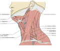 Musculi coli base.svg 1.150×975; 130 kB
-
-

-
-
 P9010880deppa.jpg 2.560×1.920; 2,53 MB
-
 Porsmose Man.jpg 1.920×2.560; 1,41 MB
-
 Porsmose-Man.jpg 1.571×2.095; 1,4 MB
-
 Posseltb.png 566×582; 116 kB
-
 Processus alveolaris.png 686×519; 274 kB
-
 Processus styloideus.png 1.632×1.224; 2,61 MB
-
 Processuscoronoideusmandibulae.PNG 600×402; 24 kB
-
 Processuszygomaticusossisfrontalis.PNG 450×435; 69 kB
-
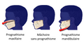 Prognathism3-fr.png 765×386; 140 kB
-
 Reduction of a dislocated mandible by a Greek surgeon. Wellcome M0013486.jpg 2.781×3.854; 3,79 MB
-
 Reproducción del cráneo y mándibula del Hombre de Grimaldi.jpg 3.632×3.660; 6,88 MB
-
-

-
 Restos de Homo sapiens. Museu de Prehistòria de València 03.jpg 3.288×2.511; 3,52 MB
-
 Rotation mandible bone.gif 600×600; 3,51 MB
-
 Siebert 12 (jaws).jpg 1.302×1.667; 558 kB
-
 Siebert 12.jpg 1.946×2.854; 1,33 MB
-
 Skull reconstruction of Australopithecus sediba.jpg 8.034×4.854; 3,34 MB
-
 Skullfront.png 1.220×1.872; 1,81 MB
-
 Slide10dddd.JPG 960×720; 102 kB
-
 Slide2tttt.JPG 960×720; 46 kB
-
 Slide3tttt.JPG 960×720; 54 kB
-
 Slide3yyyy.JPG 960×720; 111 kB
-
 Slide7oooo.JPG 960×720; 78 kB
-
 Sobo 1906 331.png 1.647×1.050; 4,96 MB
-
 Sobo 1906 338.png 1.512×1.419; 2,05 MB
-
 Sobo 1906 340.png 1.878×654; 1,17 MB
-
 Sobo 1906 341.png 1.329×930; 1,18 MB
-
 Sobo 1906 348.png 1.362×897; 1,17 MB
-
 Sobo 1906 349.png 1.086×777; 826 kB
-
 Sobo 1906 359.png 1.875×1.005; 5,4 MB
-
 Sobo 1909 90.png 2.044×1.276; 7,48 MB
-
 Sobo 1909 91.png 2.220×1.468; 9,34 MB
-
 Sobo 1909 92.png 2.092×1.664; 588 kB
-
 Sobo 1909 93.png 1.764×1.236; 6,25 MB
-
 Solutrense de la Cueva del Parpalló 02.jpg 4.000×2.667; 4,82 MB
-
 Sotokawazu01.jpg 505×435; 46 kB
-
 Sotokawazu02.jpg 482×438; 39 kB
-
 Sotokawazu03.jpg 550×378; 43 kB
-
 Sotokawazu29.jpg 503×488; 51 kB
-
 Sotokawazu30.jpg 485×429; 52 kB
-
 Sotokawazu31A.jpg 496×410; 47 kB
-
 Sotokawazu32A.jpg 510×434; 46 kB
-
 Sotokawazu33.jpg 433×493; 49 kB
-
 Sotokawazu35.jpg 495×401; 40 kB
-
 Spalteholz's Hand-Atlas of Human Anatomy (1906) - Vol 1 - Fig 045.png 2.072×1.768; 671 kB


.jpg)
.jpg)
.jpg)
.jpg)
.jpg)

_-_Fig_166.png)
_-_Fig_403.png)
_-_Fig_404.png)
.jpg)

_(14592310829).jpg)


_-_Fig_110.png)
_-_Fig_111.png)
_-_Fig_112.png)


_(14771022185).jpg)
_-_Fig._234.png)
_-_Fig._235.png)
_-_Fig._237.png)


_-_Plt17_Fig01.png)
_-_Plt17_Fig02.png)
_-_Plt18_Fig01.png)
_2.JPG)


.png)


_-_Fig_073.png)
_-_Fig_074.png)
_-_Fig_188.png)
_-_Fig_189.png)

_(14571103240).jpg)
_(14571155398).jpg)
_(14777670963).jpg)


.jpg)


_-_Vol_1_-_Fig_045.png)
_-_Fig._236.png)
.jpg)