Category:Hydrogen bonding
Jump to navigation
Jump to search
form of association between an electronegative atom and a hydrogen atom attached to a second, relatively electronegative atom  | |||||
| Upload media | |||||
| Subclass of |
| ||||
|---|---|---|---|---|---|
| |||||
Subcategories
This category has the following 8 subcategories, out of 8 total.
Media in category "Hydrogen bonding"
The following 200 files are in this category, out of 202 total.
(previous page) (next page)-

-
 (NiCl2(OH2)4)(H2O)2+Hbonding.jpg 618 × 518; 53 KB
-
 2-pyridone dimer.svg 173 × 126; 16 KB
-
 2-pyridone-chemical-dimer.png 626 × 479; 7 KB
-
 2-pyridone-chemical-dimer.svg 626 × 479; 19 KB
-
 2-Pyridone-dimer-(lactam)-3D-balls.png 1,282 × 2,000; 391 KB
-
 2-Pyridone-dimer-(lactam)-3D-spacefill.png 1,410 × 2,000; 482 KB
-
 2-Pyridone-dimer-(lactim)-3D-balls.png 1,272 × 2,000; 412 KB
-
 2-Pyridone-dimer-(lactim)-3D-spacefill.png 1,401 × 2,000; 479 KB
-
 6atom int.jpg 569 × 426; 67 KB
-
 7atom int v5.jpg 512 × 453; 67 KB
-
 A minorII.png 1,385 × 975; 365 KB
-
 A-minor-motif type0.png 844 × 866; 117 KB
-
 A-minor-motif type1.png 640 × 480; 66 KB
-

-
 Acqua legami a idrogeno.jpg 917 × 971; 56 KB
-
 Allylic strain and hydrogen bonding.png 659 × 345; 6 KB
-
 Alpha helix.png 287 × 666; 129 KB
-
 Amide Hydrogen Bridge schematic V1.svg 509 × 156; 10 KB
-
 Amine12.png 357 × 156; 15 KB
-
 Amoniakoa hidrogeno loturak.png 351 × 62; 3 KB
-
 Amoniakoaren hidrogeno loturak 3D.jpg 360 × 202; 9 KB
-
 Amoniakoaren hidrogeno loturak.png 478 × 199; 5 KB
-
 Ascorbic acid H-bonding.svg 620 × 414; 17 KB
-
 Atombindung-C-C-mit-Schalen.png 436 × 231; 26 KB
-
 Atombindung-C-C-mit-Schalen.svg 419 × 221; 52 KB
-

-
 Auswirkung H-Brücke als Diagramm auf H-Verbindungen.png 637 × 437; 29 KB
-

-
 Beispiele zur Betrachtung der Polarität der Bindungen.svg 1,405 × 710; 721 KB
-
Bending-Twisting-Motions-and-Main-Interactions-in-Nucleoplasmin-Nuclear-Import-pone.0157162.s001.ogv 10 s, 1,146 × 684; 8.28 MB
-
Bending-Twisting-Motions-and-Main-Interactions-in-Nucleoplasmin-Nuclear-Import-pone.0157162.s002.ogv 10 s, 1,146 × 684; 8.29 MB
-
Bending-Twisting-Motions-and-Main-Interactions-in-Nucleoplasmin-Nuclear-Import-pone.0157162.s003.ogv 10 s, 1,146 × 684; 7.35 MB
-
 BetaBendRibbon.pdf 1,275 × 1,650; 85 KB
-
 BetaBendRibbon.png 294 × 105; 20 KB
-
 Bifunkcyjny katalizator tiomocznikowy.png 1,105 × 676; 11 KB
-
 BiliburinHydrogenBond.png 3,760 × 3,314; 486 KB
-
 BiliburinHydrogenBond.svg 353 × 309; 40 KB
-
 Boiling-points Chalcogen-Halogen.Ar.png 337 × 224; 14 KB
-
 Boiling-points Chalcogen-Halogen.svg 337 × 224; 23 KB
-
 Boric-acid-layer-3D-balls.png 1,000 × 611; 279 KB
-
 C-H-Bindung-fuer-Elektronegativitaet.svg 409 × 216; 28 KB
-
 Carbene radical H bonding.png 500 × 359; 21 KB
-
 Carboxylic Acids Hydrogen Bonds V.1.png 1,900 × 785; 16 KB
-
 CCAARiboseZipper.PNG 640 × 408; 111 KB
-
 CompareHB XB.png 1,471 × 385; 19 KB
-
-
-
Data-of-the-molecular-dynamics-simulations-of-mutations-in-the-human-connexin46-docking-interface-mmc2.ogv 2 min 23 s, 850 × 456; 14 MB
-
Data-of-the-molecular-dynamics-simulations-of-mutations-in-the-human-connexin46-docking-interface-mmc3.ogv 2 min 5 s, 850 × 456; 10.26 MB
-
Data-of-the-molecular-dynamics-simulations-of-mutations-in-the-human-connexin46-docking-interface-mmc4.ogv 1 min 1 s, 850 × 456; 4.36 MB
-
Data-of-the-molecular-dynamics-simulations-of-mutations-in-the-human-connexin46-docking-interface-mmc5.ogv 1 min 49 s, 850 × 456; 8.09 MB
-
Data-of-the-molecular-dynamics-simulations-of-mutations-in-the-human-connexin46-docking-interface-mmc6.ogv 2 min 31 s, 850 × 456; 16.93 MB
-
-
-
Developing-Hypothetical-Inhibition-Mechanism-of-Novel-Urea-Transporter-B-Inhibitor-srep05775-s1.ogv 30 s, 480 × 480; 6.27 MB
-
Developing-Hypothetical-Inhibition-Mechanism-of-Novel-Urea-Transporter-B-Inhibitor-srep05775-s10.ogv 31 s, 480 × 480; 6.78 MB
-
Developing-Hypothetical-Inhibition-Mechanism-of-Novel-Urea-Transporter-B-Inhibitor-srep05775-s11.ogv 36 s, 480 × 480; 7.74 MB
-
Developing-Hypothetical-Inhibition-Mechanism-of-Novel-Urea-Transporter-B-Inhibitor-srep05775-s12.ogv 30 s, 480 × 480; 6.79 MB
-
-
Developing-Hypothetical-Inhibition-Mechanism-of-Novel-Urea-Transporter-B-Inhibitor-srep05775-s3.ogv 30 s, 480 × 480; 6.23 MB
-
Developing-Hypothetical-Inhibition-Mechanism-of-Novel-Urea-Transporter-B-Inhibitor-srep05775-s4.ogv 30 s, 480 × 480; 6.34 MB
-
Developing-Hypothetical-Inhibition-Mechanism-of-Novel-Urea-Transporter-B-Inhibitor-srep05775-s5.ogv 29 s, 480 × 480; 5.94 MB
-
Developing-Hypothetical-Inhibition-Mechanism-of-Novel-Urea-Transporter-B-Inhibitor-srep05775-s6.ogv 31 s, 480 × 480; 6.68 MB
-
Developing-Hypothetical-Inhibition-Mechanism-of-Novel-Urea-Transporter-B-Inhibitor-srep05775-s7.ogv 31 s, 480 × 480; 6.62 MB
-
Developing-Hypothetical-Inhibition-Mechanism-of-Novel-Urea-Transporter-B-Inhibitor-srep05775-s8.ogv 35 s, 480 × 480; 7.83 MB
-
Developing-Hypothetical-Inhibition-Mechanism-of-Novel-Urea-Transporter-B-Inhibitor-srep05775-s9.ogv 36 s, 480 × 480; 7.89 MB
-
 DihydrogenBondingBH4-H2O.png 691 × 736; 19 KB
-
 Dimedone-hydrogen-bonded-chain-from-xtal-3D-balls.png 835 × 1,100; 253 KB
-
-
-
 DIMERO.jpg 342 × 120; 9 KB
-
 DNA base pairing.svg 512 × 512; 291 KB
-
 DNA double helix grooves.svg 512 × 512; 143 KB
-
 DNA double helix.svg 512 × 512; 43 KB
-
 EditedA-minor-motif type1.png 443 × 344; 59 KB
-
 Ethanol-xtal-1976-3D-balls.png 1,100 × 814; 413 KB
-
Exploring-the-Origin-of-Differential-Binding-Affinities-of-Human-Tubulin-Isotypes-αβII-αβIII-and-pone.0156048.s011.ogv 17 s, 1,840 × 1,056; 29.64 MB
-
Exploring-the-Origin-of-Differential-Binding-Affinities-of-Human-Tubulin-Isotypes-αβII-αβIII-and-pone.0156048.s012.ogv 17 s, 1,840 × 1,056; 34.46 MB
-

-

-
 Fluro binding model.png 385 × 272; 6 KB
-
 Formula e-Konkani Vishwakosh.png 281 × 100; 4 KB
-
 G-quadruplex.jpg 440 × 271; 30 KB
-
GFPs-Mechanical-Intermediate-States-pone.0046962.s003.ogv 1 min 9 s, 852 × 480; 23.95 MB
-
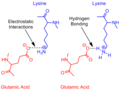
 Glutamic Acid Lysine salt bridge.png 710 × 774; 10 KB
-
 Glyoxylic acid H-bonded.png 489 × 317; 14 KB
-
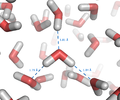
 Grotthuss mechanism.jpg 1,512 × 1,205; 126 KB
-
 H bond in RNA translation.jpg 378 × 394; 36 KB
-
 H bruck Komplexe01.jpg 4,316 × 2,413; 355 KB
-
 H-bonding alcohol.png 745 × 256; 3 KB
-
 H-bonding vs chalcogen bonding.jpg 1,092 × 948; 248 KB
-
 H-bonds of N, Q, S, T, Y.tif 1,434 × 678; 3.71 MB
-
 H-Bruecke.svg 222 × 150; 17 KB
-
 H-Brücken fast wie Bindung mit KWM erklären.svg 478 × 365; 50 KB
-
 H-donor-acceptor.svg 550 × 588; 10 KB
-
 H-O configuration conservation.gif 1,280 × 720; 103 KB
-
 H3OBF4subunit.png 981 × 937; 43 KB
-
 Hbond lengths unannotated.svg 425 × 421; 35 KB
-
 Hbond lengths.svg 425 × 421; 38 KB
-
 Hbondalcohol.png 926 × 365; 33 KB
-
 HBondApprox.png 1,890 × 531; 19 KB
-
 HF Hydrogen-bonding illust jp.svg 700 × 400; 53 KB
-
 Hidrogeno lotura Intramolekularra.jpg 283 × 177; 9 KB
-
 Hidrogeno lotura intramolekularra.png 295 × 187; 4 KB
-
 Hidrogeno lotura intramolekularrak.png 462 × 295; 8 KB
-
 Hidrogeno loturak ur molekulan.png 800 × 794; 82 KB
-
 Hydrogen Bond Quadruple AngewChemIntEd 1998 v37 p75.jpg 1,138 × 922; 235 KB
-
 Hydrogen bond stabilization.png 544 × 430; 7 KB
-
 Hydrogen bond.png 640 × 480; 38 KB
-

-

-
 Hydrogen Bondings-es.jpg 1,230 × 698; 198 KB
-
 Hydrogen bondings.svg 512 × 291; 8 KB
-
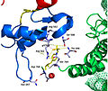 Hydrogen bonds at pY766.jpg 858 × 720; 239 KB
-

-
 Hydrogen Bonds in Baeyer-Villiger Reaction.png 778 × 594; 11 KB
-
 Hydrogen bridge.jpg 359 × 270; 18 KB
-
 Hydrogen-covalent-bond-forces-(simple).svg 446 × 329; 52 KB
-
 Hydrogenbond.jpg 640 × 512; 18 KB
-
 Hydrogenbonding.jpg 316 × 242; 25 KB
-
 HydrogenhydrogenbondingE.svg 548 × 150; 10 KB
-
 HydrophobicOrdering.jpg 367 × 435; 23 KB
-
Identification-of-Chalcones-as-Fasciola-hepatica-Cathepsin-L-Inhibitors-Using-a-Comprehensive-pntd.0004834.s009.ogv 1 min 12 s, 640 × 480; 832 KB
-
Identification-of-Chalcones-as-Fasciola-hepatica-Cathepsin-L-Inhibitors-Using-a-Comprehensive-pntd.0004834.s010.ogv 1 min 9 s, 640 × 480; 403 KB
-
Induced-Fit-in-Protein-Multimerization-The-HFBI-Case-pcbi.1005202.s004.ogv 50 s, 1,234 × 1,126; 44.56 MB
-
Induced-Fit-in-Protein-Multimerization-The-HFBI-Case-pcbi.1005202.s005.ogv 4.0 s, 1,412 × 1,030; 3.57 MB
-
 Intensitàlegameidrogeno.png 321 × 245; 4 KB
-
 Intra-H bond.png 721 × 576; 15 KB
-
 Jp-2017-09269z 0001.gif 500 × 192; 39 KB
-
 Julia-Colonna Active Site Structure1.png 1,289 × 2,121; 148 KB
-
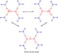 Kation Zundela.png 2,081 × 1,868; 36 KB
-
 Kevlar chemical structure H-bonds.svg 512 × 246; 19 KB
-
 Kyselina sírová - vodíkové vazby délky.jpg 5,120 × 2,642; 511 KB
-
 Lewisschreibweise mit Verdeutlichung der Wasserstoffbrücken.svg 965 × 398; 55 KB
-
 Liquid water hydrogen bond.png 1,354 × 1,124; 325 KB
-
 Major groove triples.png 847 × 393; 78 KB
-
 Mecanisme grotthuss.tif 1,512 × 1,205; 5.23 MB
-
-
-
-
-
 MEP von Propanol.png 804 × 618; 195 KB
-
 MEP von Syn-Dichlorethen.png 902 × 699; 204 KB
-
 Methanol Hydrogen Bridge V.1.svg 441 × 211; 21 KB
-
 Methanol Hydrogen Bridge V.2.svg 444 × 213; 22 KB
-
Modeling-Conformational-Ensembles-of-Slow-Functional-Motions-in-Pin1-WW-pcbi.1001015.s016.ogv 5.0 s, 948 × 894; 2.25 MB
-
-
-
-
-
-
-
-
-
-
 Mpemba HB model.gif 356 × 236; 25 KB
-
 Next Revisit Glutamic Acid Lysine salt bridge.png 1,085 × 759; 24 KB
-
 Nucleic acid polymerization.svg 512 × 512; 95 KB
-
 Nylon6,6xtal.svg 345 × 238; 56 KB
-
 O-H-Bindung-fuer-Elektronegativitaet.png 426 × 225; 19 KB
-
 O-H-Bindung-fuer-Elektronegativitaet.svg 409 × 216; 28 KB
-
 O-Nitroanilin Wasserstoffbrücke.svg 154 × 148; 10 KB
-
 O-Nitrophenol Wasserstoffbrücke.svg 148 × 148; 9 KB
-
 Oxyanion hole.png 993 × 1,067; 20 KB
-
-

-
 Quadruplex1.png 640 × 434; 48 KB
-
 Redeux Glutamic Acid Lysine salt bridge.png 1,068 × 809; 22 KB
-
 Revisited Glutamic Acid Lysine salt bridge.png 1,068 × 759; 22 KB
-
 Ricinolsäure cyclisch.svg 377 × 213; 18 KB
-
 Rizinolsäure cyclisch.svg 489 × 329; 9 KB
-
 RL nest bound to an egg oxygen atom.pdf 1,275 × 1,650; 85 KB
-
 RNA major groove triple.png 640 × 434; 72 KB
-
 Salicylaldehyd Wasserstoffbrücke.svg 128 × 158; 3 KB
-
 Salt bridge (Glutamate x Lysin) SK.png 550 × 600; 33 KB
-
 Schmelz+Siedepunkte.jpg 635 × 469; 35 KB
-
 Sdp-H-Bruecke H2X.svg 850 × 600; 95 KB
-
 Sdp-H-Bruecke-Alkanole.svg 850 × 600; 153 KB
-
-
 Sn snapshot.png 597 × 607; 153 KB
-
 Srep03005-f1.gif 100 × 122; 17 KB
-
-
 Structure of the G–G mismatch.png 702 × 361; 77 KB
-
 Tetrahedral Structure of Water-sl.png 500 × 448; 50 KB
-
 Tetrahedral Structure of Water.png 500 × 448; 64 KB
-
 Transition States hydrogen-bridged cations.jpg 255 × 157; 18 KB
-
-
-
 TriuretHbond.png 502 × 309; 14 KB
-
Ultrastable-cellulosome-adhesion-complex-tightens-under-load-ncomms6635-s2.ogv 55 s, 800 × 450; 21.91 MB
-
Ultrastable-cellulosome-adhesion-complex-tightens-under-load-ncomms6635-s3.ogv 2 min 1 s, 800 × 450; 42.59 MB
-
 Unconventional hydrogen bonding in transition metal hydrides complexes.png 1,334 × 308; 53 KB
-
 Ur molekulen arteko hidrogeno lotura.jpg 866 × 313; 22 KB
-
 Urea as a nucleic acid denaturant.svg 512 × 512; 94 KB
-
 WikipediaHDonorAcceptor.png 1,023 × 1,045; 41 KB
-
 WikipediaHDonorAcceptor.svg 244 × 249; 10 KB
-
 Wobble base pairing.svg 512 × 512; 224 KB
-
 Zeichnung zum Begriff Dipolmoment mit Pfeilen.svg 742 × 451; 35 KB
-
 Zustandekommen von H-Brücken mit KWM erklären.svg 1,455 × 332; 68 KB
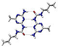
_Representative_hydrogen_bond_patterns_in_supramolecular_assembly._(b)_Hydrogen_bond_network_in_cyanuric_acid-melamine_crystals.png)
4)(H2O)2%2BHbonding.jpg)


-3D-balls.png)
-3D-spacefill.png)
-3D-balls.png)
-3D-spacefill.png)


.svg)


_SK.png)


_DNA_duplex_formation_and_(b)_protein_%CE%B2-sheet_structure.png)