Category:Hydrozoa illustrations
Jump to navigation
Jump to search
Zoological illustrations of the Hydrozoa
Subcategories
This category has the following 4 subcategories, out of 4 total.
H
T
Media in category "Hydrozoa illustrations"
The following 67 files are in this category, out of 67 total.
-
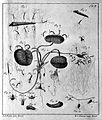
Abraham Trembley, Polyp Movement.jpg 3,946 × 5,397; 6.48 MB
-
Atti della Società italiana di scienze naturali (1876) (20322241206).jpg 2,442 × 3,500; 1.51 MB
-
Campanularia hicksoni .png 769 × 1,021; 64 KB
-
EB1911 Hydromedusae - Aglantha rosea, a British medusa.jpg 302 × 542; 65 KB
-
EB1911 Hydromedusae - Budding from the Ectoderm in Margellium.jpg 869 × 667; 143 KB
-
EB1911 Hydromedusae - calcareous corallum Millepora nodosa.jpg 377 × 419; 64 KB
-
EB1911 Hydromedusae - Carmarina hastata.jpg 650 × 806; 241 KB
-
EB1911 Hydromedusae - Clavatella prolifera - ambulatory medusa.jpg 571 × 300; 72 KB
-
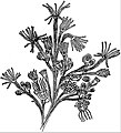
EB1911 Hydromedusae - Colony of Bougainvillea fruticosa.jpg 496 × 655; 87 KB
-
EB1911 Hydromedusae - corallum of Astylus subviridis.jpg 292 × 767; 49 KB
-
EB1911 Hydromedusae - Cunina rhododactyla.jpg 490 × 343; 55 KB
-
EB1911 Hydromedusae - development of Phialidium temporarium.jpg 850 × 288; 93 KB
-
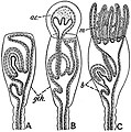
EB1911 Hydromedusae - Diphyes campanulata.jpg 508 × 1,114; 93 KB
-
EB1911 Hydromedusae - formation of an Acrocyst and a Marsupium.jpg 606 × 612; 114 KB
-
EB1911 Hydromedusae - Medusa budding with the formation of an entocodon.jpg 604 × 1,324; 179 KB
-
EB1911 Hydromedusae - modifications of a Calyptoblastic Hydromedusa.jpg 551 × 892; 147 KB
-
EB1911 Hydromedusae - modifications of a colony of Siphonophora.jpg 469 × 861; 105 KB
-
EB1911 Hydromedusae - Muscular Cells of Medusae Lizzia.jpg 594 × 286; 26 KB
-
EB1911 Hydromedusae - Ocellus of Lizzia koellikeri.jpg 276 × 520; 77 KB
-
EB1911 Hydromedusae - Octorchandra canariensis.jpg 660 × 780; 174 KB
-
EB1911 Hydromedusae - Olindias mülleri.jpg 527 × 1,090; 248 KB
-
EB1911 Hydromedusae - Oral Surface of one of the Leptomedusae.jpg 492 × 484; 78 KB
-
EB1911 Hydromedusae - Physalia anatomy.jpg 732 × 1,231; 196 KB
-
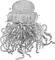
EB1911 Hydromedusae - Porpita seen from above.jpg 516 × 525; 126 KB
-
EB1911 Hydromedusae - Portion of colony of Bougainvillea fruticosa.jpg 818 × 903; 185 KB
-
EB1911 Hydromedusae - Pteronema darwinii.jpg 369 × 880; 106 KB
-
EB1911 Hydromedusae - significance of the Entocodon in Medusa-buds.jpg 597 × 1,341; 215 KB
-
EB1911 Hydromedusae - statocyst of Geryonia (Carmarina hastata).jpg 606 × 408; 123 KB
-
EB1911 Hydromedusae - Statocyst of Mitrocoma annae.jpg 476 × 458; 55 KB
-
EB1911 Hydromedusae - Statocyst of Octorchis.jpg 394 × 248; 45 KB
-
EB1911 Hydromedusae - Statocyst of Phialidium.jpg 478 × 253; 37 KB
-
EB1911 Hydromedusae - Stephalia corona - a young colony.jpg 844 × 917; 245 KB
-
EB1911 Hydromedusae - Stomotoca divisa.jpg 303 × 423; 49 KB
-
EB1911 Hydromedusae - Structure of the Gonophore.jpg 668 × 1,042; 252 KB
-
EB1911 Hydromedusae - structure of Velella.jpg 971 × 1,418; 238 KB
-
EB1911 Hydromedusae - Tentaculocyst of Cunina lativentris.jpg 515 × 492; 95 KB
-
EB1911 Hydromedusae - Tentaculocyst of Cunina solmaris.jpg 512 × 518; 92 KB
-
EB1911 Hydromedusae - Tiara pileata.jpg 598 × 1,054; 201 KB
-
EB1911 Hydromedusae - Upper surface of Velella.jpg 320 × 171; 21 KB
-
EB1911 Hydrozoa Fig. 1.JPG 835 × 498; 103 KB
-
EB1911 Hydrozoa Fig. 2.JPG 302 × 397; 42 KB
-
EB1911 Hydrozoa Fig. 3.JPG 872 × 539; 60 KB
-
EB1911 Hydrozoa Fig. 4.JPG 595 × 842; 91 KB
-
Hydromedusae23.jpg 213 × 665; 33 KB
-
Hydrozoa from J.C. Schaffer "Die armpolypen", 1763 Wellcome L0002440.jpg 1,260 × 1,492; 799 KB
-
Hydrozoa from; J.C. Schaffer "Armpolypen", 1763 Wellcome L0002442.jpg 1,246 × 1,482; 758 KB
-
Hydrozoa from; Mikroskopische Gemuths-und Augen Ergotzung Wellcome L0002486.jpg 1,212 × 1,742; 1.03 MB
-
Hydrozoa from; Mikroskopische Gemuths-und Augen Ergotzung Wellcome L0002487.jpg 1,176 × 1,552; 707 KB
-
Hydrozoa; J.C. Schaffer "Armpolypen...", 1763 Wellcome L0002441.jpg 1,264 × 1,490; 836 KB
-
Lectures on the elements of comparative anatomy (Page 21) BHL3319011.jpg 2,326 × 3,749; 637 KB
-
Medusae of world-vol02 fig135-139 Clytia hemisphaerica - Campanularia volubilis.jpg 1,634 × 1,782; 447 KB
-
Neoturris papua Cytaeis tetrastyla Aglaura hemistoma.JPG 514 × 794; 42 KB
-
PSM V16 D660 Gonophores of the hydrozoa.jpg 1,195 × 442; 94 KB
-
PTRS168 0639.png 2,445 × 3,274; 1.05 MB
-
Text-book of comparative anatomy (1898) (14778983895).jpg 1,008 × 1,288; 175 KB
-
The pedigree of man - and other essays (1903) (14762380224).jpg 1,238 × 2,140; 222 KB
-
The pedigree of man - and other essays (1903) (14762398124).jpg 1,258 × 2,168; 216 KB
-
Trembley - Mémoires pour l'histoire des polypes 96.jpg 1,952 × 2,684; 911 KB


