Category:Imaging
Jump to navigation
Jump to search
form of signal processing for which the input is an image and the output is either an image or a set of characteristics or parameters related to the image | |||||
| Upload media | |||||
| Subclass of | |||||
|---|---|---|---|---|---|
| Different from | |||||
| |||||
Subcategories
This category has the following 34 subcategories, out of 34 total.
3
A
- Imaging artifacts (5 F)
C
D
E
- Event-based imaging (5 F)
F
I
- In vivo imaging (68 F)
K
- Kinetic imaging (9 F)
M
- Multimodal imaging (12 F)
O
P
- Imaging phantoms (21 F)
- Pulmonary imaging (7 F)
Q
- Quantum imaging (5 F)
R
S
T
W
- Whole body imaging (9 F)
Media in category "Imaging"
The following 97 files are in this category, out of 97 total.
-
 1D defocused optical transfer function.svg 512 × 384; 24 KB
-
 1D diffraction limited optical transfer function..svg 512 × 384; 24 KB
-
 Arabidopsis root hairs.jpg 2,752 × 2,208; 201 KB
-
 Arteries absorption.png 633 × 485; 21 KB
-
 Astronomy career inspirations.jpg 1,600 × 1,600; 284 KB
-
 Balajinewsm.png 500 × 500; 31 KB
-
-
 Bilderzeugung.png 1,920 × 800; 48 KB
-
 Breast absorption.png 633 × 482; 21 KB
-
 Brtxum.jpg 706 × 706; 98 KB
-
 Calcium in Pollen.jpg 2,048 × 2,048; 931 KB
-
 Case-3-mri.png 813 × 615; 368 KB
-
 Central projection of a real camera - no annotation.svg 833 × 429; 15 KB
-
 Central projection of a real camera.svg 833 × 429; 22 KB
-
 CHP mouse heart section.jpg 935 × 1,071; 278 KB
-
 Cone-resolved visual acuity testing.gif 442 × 221; 14.41 MB
-
 Confocal Raman Image of a pharmaceutical emulsion..png 2,048 × 2,048; 5.21 MB
-
 Convvex mirror1-Hebrew.jpg 934 × 705; 63 KB
-

-
 Definitions PSF OTF MTF PhTF.svg 1,065 × 568; 767 KB
-
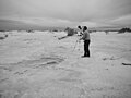
 Documenting the Tracks from a Different View (8c889a0d-3cce-45dd-a05a-2f37c6e1ad67).JPG 4,389 × 2,784; 3.2 MB
-
 DOF palette-2019-07-03-Gianmaria Calisesi.png 1,972 × 662; 164 KB
-
 DTschematic.jpg 578 × 616; 44 KB
-
 Ellipsoid eigenvalues14142.jpg 560 × 420; 20 KB
-
 Ellipsoid2722.jpg 560 × 420; 21 KB
-
 Ellipsoid888.jpg 560 × 420; 24 KB
-
 Energy gap lax rouge.jpg 493 × 429; 28 KB
-
 FluorescenceImaging.JPG 3,008 × 2,000; 1.8 MB
-
 Foxtenn cams.jpg 1,140 × 685; 831 KB
-
 Framesovertime.png 383 × 262; 14 KB
-
 FRET Jablonski diagram.svg 733 × 715; 208 KB
-
 Grypaploskyrovskiy.jpg 1,800 × 1,200; 141 KB
-
 Guambra Más Linda de Riobamba.jpg 1,920 × 2,560; 460 KB
-

-
 Ht1 con02.png 457 × 411; 116 KB
-
 Ibrahimsamake.jpg 800 × 800; 138 KB
-
 IbrahimSamakeParisienne.jpg 3,024 × 4,032; 3.2 MB
-
 IECCD55-20.jpg 2,223 × 1,457; 1.08 MB
-
 Illum room.jpg 1,782 × 1,036; 73 KB
-

-

-
 Ilogo94.jpg 2,463 × 1,905; 274 KB
-
 Image1 dinnumammu.jpg 3,024 × 4,032; 2.04 MB
-
 Imaging out of box.jpg 225 × 225; 5 KB
-
 IMG-20230704-WA0005.jpg 2,048 × 1,532; 185 KB
-
 IMG-20230704-WA0007.jpg 1,364 × 1,023; 269 KB
-
 Intact antibodies and a variety of antibody fragments.png 942 × 806; 601 KB
-
 IVIS imaging of murine tumours.jpg 1,956 × 1,371; 327 KB
-
 Kaktus logo.jpg 456 × 210; 32 KB
-
 Laboratory edge-illumination system 04.png 3,311 × 2,148; 412 KB
-
 Long wave radiaton.jpg 1,008 × 630; 94 KB
-
 MALDI image of rat lung.jpg 766 × 2,146; 253 KB
-
 MALDI imaging.png 2,664 × 1,453; 1.01 MB
-
 Miladkanka.jpg 1,440 × 1,440; 202 KB
-
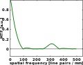 MTF of an optical system with spherical aberration..svg 512 × 402; 28 KB
-
 Optische Abbildungsgeometrie einer Kamera.svg 833 × 549; 51 KB
-
 OTF of an optical system with spherical aberration..svg 512 × 409; 29 KB
-
 Out-of-focus image of a spoke target..svg 512 × 512; 424 KB
-
 Pasig City Bike Share ADB San Antonio Escriva Drive Ortigas Center.jpg 4,032 × 2,268; 4.52 MB
-
 Perfil (1) Pablo Santoro.jpg 1,080 × 1,080; 871 KB
-
 PH-responsive nanogel for imaging.png 1,726 × 498; 461 KB
-
 Phase contrast imaging schematic.png 1,526 × 803; 103 KB
-
 PNI RF Pulse Sequencing.png 343 × 199; 11 KB
-
 Positron emission tomography.png 1,050 × 736; 531 KB
-
 Principles of imaging geometry of a camera no annotation.svg 833 × 549; 42 KB
-
 Principles of imaging geometry of a camera.svg 833 × 549; 53 KB
-
 Prinzip.Bildgebung.durch.Lichtwandlung.png 2,048 × 2,048; 60 KB
-
 Rdhard Oficial.jpg 1,254 × 1,254; 80 KB
-
 Reale Zentralprojektion einer Kamera.svg 833 × 429; 22 KB
-
 Sarfus PolarisationState.jpg 552 × 313; 22 KB
-
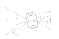
 Schematic view of imaging and diffraction modes in TEM..tif 4,079 × 3,654; 42.64 MB
-
 SF9 Cells phase contrast Lara Rudman.jpg 1,800 × 1,350; 459 KB
-
 Shangri-La Plaza Bike Parking.jpg 2,268 × 4,032; 7.01 MB
-
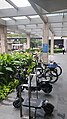
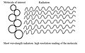 Short wave radiation.jpg 1,008 × 630; 121 KB
-
 Sindh calcture.jpg 452 × 678; 63 KB
-
-
 Skybox Imaging.jpg 5,183 × 3,456; 8.99 MB
-
 SM East Ortigas Bike Parking.jpg 3,600 × 2,400; 2.1 MB
-
 SM Megamall Bike Parking.jpg 4,032 × 3,024; 8.35 MB
-
 Spessore.jpg 665 × 298; 13 KB
-
 Spoke target imaged with spherical aberration.svg 512 × 512; 334 KB
-
 SuperResolutionMulticolorStrategies.png 638 × 442; 11 KB
-
 Tampak Depan Kantor Desa Patanyamang.jpg 714 × 402; 99 KB
-
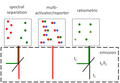
 Teerahertz near-field array for μm-scale surface imaging (cropped).png 768 × 257; 343 KB
-
 Teerahertz near-field array for μm-scale surface imaging.png 864 × 1,072; 1.14 MB
-
 The Lions Gate (1917341250).jpg 906 × 864; 530 KB
-
 There is no such turban anywhere in the world except in Sindh.jpg 518 × 690; 70 KB
-
 Through the 3D Lens (4b34bc6c-1234-4e63-8f46-a1116ea36bc2).JPG 4,592 × 3,448; 4.63 MB
-
 Toad Bug.jpg 2,158 × 1,429; 1.01 MB
-
 Tomato root hairs.jpg 2,752 × 2,208; 423 KB
-
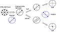
 Trefoil aberration PSF OTF and example image.svg 892 × 282; 1.21 MB
-
 University of Santo Tomas Bike Parking.jpg 2,268 × 3,024; 1.79 MB
-
 Vergence-Accommodation Conflict Diagram.png 2,000 × 1,500; 246 KB
-
 Whole body by Mass Spectrometry Imaging.jpg 1,089 × 464; 106 KB
-
 کتاب اران نامه،.jpg 1,544 × 2,430; 709 KB
-
 کتاب اران نامه۰.jpg 1,685 × 2,623; 882 KB
-
 김승현 대표.jpg 5,040 × 3,360; 6.08 MB


.jpg)

.JPG)


_Pablo_Santoro.jpg)


.jpg)

.JPG)


.png)