Category:Kidneys
Jump to navigation
Jump to search
Afrikaans: Nier
· العربية: كلية
· Aragonés: Riñón
· Azərbaycanca: Böyrəklər
· Bahasa Indonesia: Ginjal
· Bahasa Melayu: Buah pinggang
· Jawa: Ginjel
· Sunda: Ginjal
· বাংলা: বৃক্ক
· Bosanski: Bubreg
· Български: Бъбрек
· Català: Ronyó
· Čeština: Ledvina
· Cymraeg: Aren
· Dansk: Nyre
· Deutsch: Niere
· Eesti: Neerud
· English: Kidney
· Español: Riñón
· Esperanto: Reno
· Euskara: Giltzurrun
· فارسی: کلیه
· Français : Rein
· Galego: Ril
· 한국어: 콩팥
· Hrvatski: Bubreg
· Íslenska: Nýra
· Italiano: Rene
· עברית: כליה
· Kiswahili: Figo
· Kurdî: Gurçik
· Latina: Ren
· Latviešu: Nieres
· Lietuvių: Inkstas
· Magyar: Vese
· Македонски: Бубрег
· മലയാളം: വൃക്ക
· Nederlands: Nier
· 日本語: 腎臓
· Norsk bokmål: Nyre
· Norsk nynorsk: Nyre
· Oromoo: Kalee
· Kapampangan: Batu
· Polski: Nerka
· Português: Rim
· Română: Rinichi
· Runa Simi: Wasa ruru
· Русский: Почка
· Scots: Neer
· Sicilianu: Rini
· Slovenčina: Oblička
· Slovenščina: Ledvica
· Српски / srpski: Бубрег
· Srpskohrvatski / српскохрватски: Bubreg
· Suomi: Munuainen
· Svenska: Njure
· Tagalog: Bato
· தமிழ்: சிறுநீரகம்
· తెలుగు: మూత్రపిండము
· ไทย: ไต
· Türkçe: Böbrek
· اردو: كُلیَہ
· Võro: Rauh
· ייִדיש: ניר
· Українська: Нирки
· Чӑвашла: Пӳре
· 中文:腎
· internal organ in most animals, including vertebrates and some invertebrates  | |||||
| Upload media | |||||
| Pronunciation audio | |||||
|---|---|---|---|---|---|
| Instance of |
| ||||
| Subclass of |
| ||||
| Part of | |||||
| Has part(s) | |||||
| Different from | |||||
| |||||
Subcategories
This category has the following 9 subcategories, out of 9 total.
A
C
H
R
S
V
Media in category "Kidneys"
The following 50 files are in this category, out of 50 total.
-
 A stained kidney print from a fish infected by Renibacterium salmoninarum.jpg 1,300 × 1,030; 287 KB
-
 ANATOMÍA DEL RIÑÓN.jpg 5,312 × 2,988; 5.26 MB
-
 ANATOMÍA RENAL.jpg 2,959 × 2,392; 5.97 MB
-
 Atv3.jpg 571 × 612; 71 KB
-
 Axial CT scan of left renal cortical cyst.png 512 × 512; 211 KB
-
 Budowa Nerki 3sp 2022.png 12,421 × 8,976; 40.23 MB
-
 CarbAnh.png 6,600 × 5,100; 2.78 MB
-
 CarbAnh2.png 3,819 × 3,601; 2.38 MB
-
 CarbAnh3.png 6,600 × 5,100; 3.22 MB
-
 CarbAnh4.png 3,819 × 3,601; 2.46 MB
-
 Chicken's Kidney.pdf 2,000 × 1,125; 1.85 MB
-
 Chickenkidneys.png 3,024 × 4,032; 15.47 MB
-
 Circulacionrenal.jpg 700 × 1,700; 198 KB
-
De-Niere.ogg 0.9 s; 17 KB
-
 Dihydrogen phosphate.jpg 395 × 435; 26 KB
-
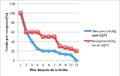 Efecte de l'AQP3 en la recuperació d'una ferida..png 472 × 299; 22 KB
-
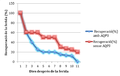 Efecte de l'AQP3 en la recuperació d'una ferida.png 578 × 364; 35 KB
-
 Egér vese (Mouse kidney).JPG 2,856 × 2,744; 1.63 MB
-
 Examples of Signs and Symptoms of Kidney Ischemia.png 720 × 504; 209 KB
-
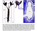 Fish kidney.jpg 931 × 775; 138 KB
-
 Fluid Flow in the Bowman's Capsule and Glomerulus.svg 512 × 384; 152 KB
-
 Glomerulus Diameter Measurement.jpg 501 × 168; 24 KB
-
 Gray1128 vie.png 624 × 614; 78 KB
-
 H λειτουργία του εγγύς εσπειραμένου σωληναρίου.png 862 × 633; 90 KB
-
 HE cystic renal dysplasia.jpg 2,448 × 1,920; 2.63 MB
-

-
 Kidney for transplant from live donor.jpg 2,560 × 1,920; 924 KB
-

-
 Nephron in kidney.png 1,920 × 1,357; 223 KB
-
 Paediatric kidney measurements.png 6,913 × 2,588; 30.09 MB
-
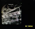 Paediatric left kidney sagittal view.jpg 3,456 × 2,911; 3.04 MB
-
 Right extrarenal pelvis as shown in axial view on ultrasound.jpg 512 × 688; 64 KB
-
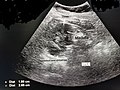
 Right extrarenal pelvis as shown in sagittal view on ultrasound.jpg 1,024 × 698; 72 KB
-
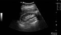 Right kidney seen on abdominal ultrasound.jpg 1,552 × 904; 323 KB
-
 Right kidney sinus fat casting posterior shadowing on ultrasound.jpg 1,024 × 698; 167 KB
-
 So that others may live 140124-Z-ZT651-003.jpg 4,928 × 3,264; 12.37 MB
-
 Stirling schematic normal renal corpuscle 2022.jpg 998 × 2,910; 1.13 MB
-
 Structure of glomerulus.png 1,024 × 1,361; 288 KB
-
 Structure of the Capillaries of the Glomerulus.jpg 1,278 × 720; 388 KB
-
 The examination of the urine of the horse and man (1911) (14770112201).jpg 1,624 × 1,928; 362 KB
-
 Three anatomical figures from Tibet Wellcome V0036134.jpg 2,691 × 3,215; 3.36 MB
-
 Tubulocolector.jpg 1,490 × 1,332; 312 KB
-
-
 Ultrasound showing hypoplastic right kidney.png 1,024 × 768; 285 KB
-
 Ultrasound showing medullary nephrocalcinosis of bilateral kidneys.png 800 × 600; 426 KB
-

-
 Yuxtaglomerulares.jpg 680 × 440; 87 KB
-
 Подоцин белок.png 1,715 × 1,264; 73 KB
-
 Эффективный циркуляционный объем31.jpg 1,268 × 1,938; 492 KB
-
 మూత్రాంగము, మూత్రపిండము, (Kidney) బొమ్మ.png 1,823 × 1,248; 434 KB


.JPG)


.jpg)


_(14770112201).jpg)


_%E0%B0%AC%E0%B1%8A%E0%B0%AE%E0%B1%8D%E0%B0%AE.png)