Category:Lichen anatomy and morphology
Jump to navigation
Jump to search
Subcategories
This category has the following 4 subcategories, out of 4 total.
Media in category "Lichen anatomy and morphology"
The following 76 files are in this category, out of 76 total.
-

-
 05 02 interactions green alga (green), fungal hypha, lichen, Lichenes (M. Piepenbring).png 3,070 × 2,302; 2.08 MB
-
 05 05 formas de crecimiento de liquenes, Lichenes (M. Piepenbring).png 3,070 × 2,302; 2.7 MB
-
 05 05 growth forms of lichens, Lichenes (M. Piepenbring).png 3,070 × 2,302; 2.71 MB
-

-
 05 09 anatomía del talo, alga verde, cianobacterias, liquenes, Lichenes (M. Piepenbring).png 3,070 × 2,302; 1.47 MB
-
 05 10 anatomy of the thallus, details, lichens, Lichenes (M. Piepenbring).png 3,070 × 2,302; 4.81 MB
-
 05 10 anatomía del talo, detalles, liquenes, Lichenes (M. Piepenbring).png 3,070 × 2,302; 4.81 MB
-
 05 11 asci and ascospores of lichens, Lichenes (M. Piepenbring).png 3,070 × 2,302; 2.38 MB
-
 05 11 ascos y ascosporas de liquenes, Lichenes (M. Piepenbring).png 3,070 × 2,302; 2.38 MB
-

-

-

-

-

-
 Abb4.10 Lichenes lichens thallus anatomy heteromerous homoiomerous 2021 (M. Piepenbring).png 3,000 × 2,250; 1.32 MB
-

-

-

-

-

-

-
-
 Abb4.54 Lichenes lichen Strigula ascus ascospores 2021 (M. Piepenbring).png 3,000 × 2,250; 1.04 MB
-
 Algal Cells, Division Ascomycota, Ascocarps, Hyphae Stained Microscope Slide.jpg 1,024 × 768; 280 KB
-
 Asco1003.jpg 1,200 × 886; 177 KB
-
 Asco1003L.jpg 1,200 × 886; 175 KB
-
 Asco1004.jpg 1,200 × 886; 244 KB
-
 Asco1004L.jpg 1,200 × 886; 242 KB
-
 Asco1005.jpg 1,200 × 886; 122 KB
-
 Asco1005L.jpg 1,200 × 886; 120 KB
-
 Asco1006.jpg 1,200 × 886; 218 KB
-
 Asco1006L.jpg 1,200 × 886; 216 KB
-
 Asco1007.jpg 1,200 × 886; 284 KB
-
 Asco1007L.jpg 1,200 × 886; 281 KB
-
 Asco1008.jpg 1,200 × 886; 326 KB
-
 Asco1008L.jpg 1,200 × 886; 324 KB
-
 Ascocarp2 Dutch text.png 666 × 600; 208 KB
-
 Bipartite and tripartite cyanolichens (10.3897-mycokeys.6.3869) Figure 1.jpg 1,512 × 1,047; 1.74 MB
-
 Brockhaus and Efron Encyclopedic Dictionary b34 867-1.jpg 1,630 × 2,747; 1.08 MB
-
 Budowa apotecjum porostów.jpg 1,969 × 960; 483 KB
-
 Cifela illustr.png 1,169 × 643; 573 KB
-
 Deyrolle 948499.jpg 2,335 × 3,113; 2.17 MB
-
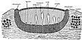
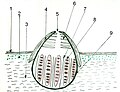
 EB1911 Lichens - apothecium and surrounding thallus.jpg 843 × 411; 180 KB
-
 EB1911 Lichens - Collema crispum.jpg 306 × 841; 65 KB
-
 EB1911 Lichens - Cora pavonia.jpg 493 × 492; 99 KB
-
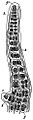 EB1911 Lichens - Ephebe pubescens.jpg 268 × 974; 118 KB
-
 EB1911 Lichens - Gyrophora cylindrica.jpg 696 × 321; 91 KB
-
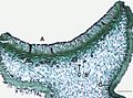
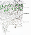
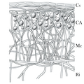
 EB1911 Lichens - heteromerous lichen thallus.jpg 463 × 806; 259 KB
-
 EB1911 Lichens - lichen-forming algae.jpg 995 × 787; 270 KB
-
 EB1911 Lichens - Usnea barbata (3).jpg 579 × 181; 50 KB
-
 EB1911 Lichens - Xanthoria parietina - apothecium.jpg 489 × 497; 99 KB
-
 File-Meyers b6 s0351a.jpg 367 × 458; 80 KB
-
 FR Lichen cross-section.png 1,712 × 1,914; 3.56 MB
-
 Intercellular transport of nutrients within the thallus of lichens.png 1,539 × 884; 467 KB
-
 Lichen Cross Section Diagram.svg 512 × 282; 94 KB
-
 Lichen cross section – heteromeric thallus.svg 516 × 532; 84 KB
-
 Lichen cross-section.png 1,712 × 1,914; 3.1 MB
-
 Lichen Diagram.svg 512 × 225; 201 KB
-
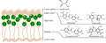 Lichen secondary metabolites.png 2,006 × 812; 441 KB
-
 Lichenographia Universalis Tab 17.jpg 3,008 × 3,560; 5.86 MB
-
 Meyers b6 s0351.jpg 800 × 1,275; 377 KB
-
 Meyers b6 s0352.jpg 800 × 1,275; 398 KB
-
 Meyers b6 s0353.jpg 800 × 1,275; 376 KB
-
 Meyers b6 s0354.jpg 800 × 1,275; 458 KB
-
 Meyers b6 s0355.jpg 800 × 1,275; 474 KB
-
 Organización de los talos heterómeros y homómeros en líquenes.png 2,000 × 973; 1.69 MB
-
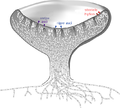
 Organización de un talo crustáceo en líquenes.png 1,100 × 1,100; 1.05 MB
-
 Organización de un talo foliáceo en líquenes.png 1,100 × 1,169; 1.38 MB
-
 Organización de un talo fruticuloso plano en líquenes.png 1,100 × 1,100; 1.22 MB
-
 Perytecjum a1.jpg 786 × 600; 143 KB
-
 Pseudocyphellen bij gewoon schildmos (Parmelia sulcata).jpg 1,372 × 967; 759 KB
-
 Rodzaje plech porostów.png 1,707 × 1,280; 843 KB
-
 Solorina saccata Sporen.jpg 1,519 × 1,139; 199 KB
-
 Лишайник в разрезе обычное освещение под микроскопом.jpg 4,912 × 3,264; 5.45 MB
-
 Лишайник пармелия в разрезе Флуоресценция под микроскопом.JPG 4,912 × 3,264; 12.58 MB
,_hifa_de_hongo,_liquenes,_Lichenes_(M._Piepenbring).png)
,_fungal_hypha,_lichen,_Lichenes_(M._Piepenbring).png)
.png)
.png)
.png)
.png)
.png)
.png)
.png)
.png)
_158.svg)
_159.svg)
_160.svg)
.svg)
.svg)
.png)
.png)
.svg)
.png)
.png)
.svg)
.png)
.svg)
.png)


_Figure_1.jpg)


.jpg)


.jpg)