Category:Mammal digestive system at the Museum of Veterinary Anatomy FMVZ USP
Jump to navigation
Jump to search
Media in category "Mammal digestive system at the Museum of Veterinary Anatomy FMVZ USP"
The following 43 files are in this category, out of 43 total.
-
 Bovine duodenal papilla.jpg 2,426 × 3,624; 3.2 MB
-
 Bovine liver.jpg 3,478 × 2,328; 4.6 MB
-
 Bovine pancreas excretory system.jpg 2,217 × 3,312; 4.19 MB
-
 Capybara liver (bile ducts).jpg 2,366 × 3,534; 3.69 MB
-
 Cat liver with gallbladder 01.jpg 2,432 × 3,633; 4.13 MB
-
 Cat liver with gallbladder 02.jpg 2,315 × 3,458; 3.43 MB
-
 Cat liver with gallbladder 03.jpg 2,475 × 3,697; 3.9 MB
-
 Cat liver. Double Gallbladder - Anomaly.jpg 2,202 × 3,289; 2.87 MB
-
 Cat small intestine.jpg 2,366 × 3,534; 3.41 MB
-
 Coati liver (bile ducts).jpg 2,190 × 3,271; 3.21 MB
-
 Coati liver.jpg 2,341 × 3,497; 3.26 MB
-
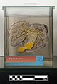
 Didactic model of a bovine omasum and abomasum-FMVZ USP-26.jpeg 2,592 × 3,872; 4.48 MB
-
 Didactic model of a bovine Rumen and Reticulum-FMVZ USP-27.jpeg 3,872 × 2,592; 4.16 MB
-
 Dog digestive tract.jpg 2,381 × 3,557; 4.04 MB
-
 Dog ilium.jpg 2,356 × 3,519; 3.25 MB
-
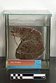
 Dog liver.jpg 3,863 × 2,586; 4.67 MB
-
 Dog spleen.jpg 2,447 × 3,655; 3.92 MB
-
 Dog stomach (open, inner view).jpg 2,354 × 3,516; 2.98 MB
-
 Dog stomach.jpg 2,473 × 3,694; 3.17 MB
-
 Domestic pig liver.jpg 1,868 × 2,790; 2.54 MB
-
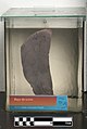 Domestic pig spleen.jpg 2,145 × 3,204; 3 MB
-
 Donkey Spleen.jpg 2,309 × 3,449; 3.77 MB
-
 Equine colon.jpg 2,277 × 3,401; 3.27 MB
-
 Equine liver.jpg 2,270 × 2,500; 3.11 MB
-
 Equine stomach-FMVZ USP-25.jpeg 3,872 × 2,592; 5.46 MB
-
 Goat liver (bile ducts).jpg 2,129 × 3,180; 3.44 MB
-
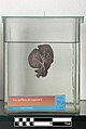
 Kangaroo liver.jpg 2,369 × 3,539; 3.52 MB
-
 Kangaroo spleen.jpg 2,592 × 3,872; 4.52 MB
-
 Kidneys and spleen of lion stillborn.jpg 2,345 × 3,503; 3.69 MB
-
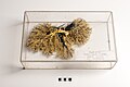
 Lion tongue and larynx.jpg 2,592 × 3,872; 4.14 MB
-
 Lynx liver.jpg 2,293 × 3,425; 3.68 MB
-
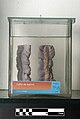
 Mule pancreas excretory system.jpg 2,442 × 3,648; 4.21 MB
-
 Nutria liver.jpg 2,150 × 3,212; 2.94 MB
-
 Ocelot liver.jpg 2,158 × 3,224; 3.07 MB
-
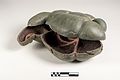
 Ovine stomach.jpg 3,233 × 2,457; 2.73 MB
-
 Rabbit stomach.jpg 3,150 × 2,109; 2.38 MB
-
 Sheep liver (bile ducts).jpg 2,267 × 3,386; 3.54 MB
-
 Sheep liver (portal vein).jpg 2,360 × 3,525; 3.97 MB
-
 Technique of formalin fixation applied to dog tongue.jpg 2,143 × 3,201; 2.54 MB
-
 Tongue of goat.jpg 3,564 × 2,386; 3.07 MB
-
 Whale Tongue.jpg 3,872 × 2,592; 5.07 MB
-
 Wistar rat digestive system.jpg 3,000 × 2,008; 1.62 MB
-
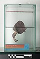
 Zebra spleen.jpg 2,378 × 3,552; 3.87 MB


.jpg)


.jpg)


.jpg)


.jpg)


.jpg)
.jpg)


