Category:Mass spectrometry
Перейти до навігації
Перейти до пошуку
analytical technique based on determining mass to charge ratio of ions  | |||||
| Завантажити медіафайл | |||||
| Є одним із |
| ||||
|---|---|---|---|---|---|
| Є підкласом |
| ||||
| Складники | |||||
| |||||
References[ред.]
- US patent application 2341551, Jr Herbert Hoover, "Mass spectrometer", published 1944-02-15 , assigned to Consolidated Engineering Co Inc
Підкатегорії
Показано 23 підкатегорії з 23.
A
- Aerosol mass spectrometry (2 F)
C
- Calutron (30 F)
I
M
- Mass spectrometry papers (38 F)
- Mass-to-charge ratio (6 F)
- Microchannel plates (9 F)
O
- Orbitrap (7 F)
P
Q
- Quadrupole mass analyzer (15 F)
S
T
- Tandem mass spectrometry (2 F)
- Time-of-flight mass spectrometry (16 F)
V
Сторінки в категорії «Mass spectrometry»
Показано 1 сторінку цієї категорії (із 1).
Файли в категорії «Mass spectrometry»
Показано 186 файлів цієї категорії (із 186).
-
 595768.fig.003a.jpg 600 × 235; 40 КБ
-
 Agilent APCI annotated.jpg 3569 × 2841; 1,69 МБ
-
 Agilent APCI sprayNeedle Annotated.jpg 3177 × 3185; 1,42 МБ
-
 Agilent APCI sprayNeedle.jpg 3177 × 3185; 1,32 МБ
-
 Agilent APCI.jpg 3093 × 2849; 1,42 МБ
-
 Agilent MixedMode annotated.jpg 3456 × 4608; 2,26 МБ
-
 Agilent MixedMode.jpg 3581 × 2773; 1,62 МБ
-
 Alignment of nanospray.jpg 4608 × 3072; 5,77 МБ
-
 APCI Source With Heated Nebulizer.png 851 × 850; 25 КБ
-
 APCI Source With LC Inlet.png 1307 × 534; 20 КБ
-
 APLI solvent cross sections de.png 677 × 921; 96 КБ
-
 Appareillage de l'IMS.png 819 × 460; 84 КБ
-
 Appareillage de la spectroscopie à mobilité ionique.png 819 × 460; 31 КБ
-
 ATCC 10876 OpenTyper openSUSE Cinnamon.png 1920 × 1054; 246 КБ
-
 Atmospheric pressure chemical ionization chamber.jpg 2712 × 2034; 952 КБ
-
 Atmospheric pressure chemical ionization.jpg 560 × 676; 42 КБ
-
 ATOFMS.png 1088 × 724; 43 КБ
-
 Basic ion funnel schematic.jpg 1366 × 768; 111 КБ
-
 Bio Threats- FDA's A-Team (6304) (9806908675).jpg 2784 × 1848; 3,8 МБ
-
 Biological Mass Spectrometry (50341723222).png 3000 × 1688; 6,1 МБ
-
 Block diagram of a mass spectrometer.jpg 1280 × 720; 138 КБ
-
 Bruker Amazon Speed ETD.jpg 1280 × 853; 117 КБ
-
 Butyric acid frag.png 822 × 624; 14 КБ
-
 Canal rays -kn.svg 1000 × 617; 5 КБ
-
 Canal rays as.svg 1000 × 617; 2 КБ
-
 Canal rays-bn.svg 1000 × 617; 2 КБ
-
 Canal rays-gu.svg 1000 × 617; 4 КБ
-
 Canal rays-mr.svg 1000 × 617; 6 КБ
-
 Canal rays-pa.svg 1000 × 617; 4 КБ
-
 Canal rays-te.svg 1000 × 617; 4 КБ
-
 Canal rays.png 800 × 491; 12 КБ
-
 Canal rays.svg 1000 × 617; 3 КБ
-
 Cellule-ICR-cubique.png 829 × 629; 17 КБ
-
 Charged.jpg 522 × 51; 4 КБ
-
 CP-4900 4chan with DMD.jpg 2505 × 1113; 1,09 МБ
-
 DDA MS Proteomics Scheme.png 907 × 491; 45 КБ
-
 DESI-IMS-TOF.png 921 × 324; 73 КБ
-
 Diagram of first representation of thermospray vaporizer.png 840 × 479; 138 КБ
-
 Diagram of fourth representation of thermospray vaporizer.png 896 × 611; 115 КБ
-
 Diagram of second representation of thermospray vaporizer.png 963 × 588; 159 КБ
-
 Diagram of third representation of thermospray vaporizer.png 922 × 473; 80 КБ
-
 Diagramme-stabilite-quadripole.png 767 × 394; 11 КБ
-
 DIOS mass spectrometry.jpg 1052 × 890; 562 КБ
-
 Direct Injection nebulizer.png 686 × 437; 36 КБ
-
 Discrete and Continuous Dynode Systems.jpg 5100 × 3300; 1,72 МБ
-
 Dual linear ion trap.JPG 3344 × 2144; 1,07 МБ
-
 ECD Sub P by DIT.jpg 843 × 465; 51 КБ
-
 Electron ionization GC-MS.png 1422 × 662; 85 КБ
-
 Electrostatic analyzer.png 960 × 720; 21 КБ
-
 Elektronspray.jpg 874 × 489; 94 КБ
-
 ESTASI scheme.tif 1890 × 848; 4,61 МБ
-
 ETD cartoon.tiff 947 × 247; 63 КБ
-
 ETD Fragmentation.tiff 782 × 161; 44 КБ
-
 ETD reaction.tiff 930 × 250; 62 КБ
-
 Faraday Cup for Plasma Diagnostics.tif 1013 × 831; 170 КБ
-
 Fast atom bombardment diagram.png 1114 × 892; 442 КБ
-
 FDA Center for Food Safety & Applied Nutrition (CFSAN) 6486 (8755007149).jpg 2784 × 1848; 3,24 МБ
-
 FDA Center for Food Safety & Applied Nutrition (CFSAN) 6487 (8755005115).jpg 1848 × 2784; 3,08 МБ
-
 FDA Center for Food Safety & Applied Nutrition (CFSAN) 6489 (8755003029).jpg 2486 × 1650; 2,34 МБ
-
 FDA Center for Food Safety & Applied Nutrition (CFSAN) 6492 (8756122836).jpg 1848 × 2784; 3,05 МБ
-
 FDA Center for Food Safety & Applied Nutrition (CFSAN) 6496 (8756119526).jpg 2600 × 1726; 2,91 МБ
-
 FDA Center for Food Safety & Applied Nutrition (CFSAN) 6498 (8756116078).jpg 2482 × 1647; 2,63 МБ
-
 Field (in-situ) spectrometrist.svg 916 × 635; 537 КБ
-
 Figure 1, Mass spectrometric analysis of eugleophycin.png 512 × 289; 35 КБ
-
 Formula para el numero de masa.png 1345 × 307; 13 КБ
-
 FT-ICR Mass spectrometer Quadrupole.jpg 4000 × 3000; 2,2 МБ
-
 FTICR cell.png 715 × 770; 120 КБ
-
 Gall' 2007 3rdRMSO Congress.jpg 513 × 700; 114 КБ
-

-
 Geschwindigkeitsfilter Kraeftegleichgewicht.svg 300 × 350; 32 КБ
-
 HDX-MS workflow.svg 301 × 939; 204 КБ
-
 Hexanal frag 1.jpg 807 × 571; 37 КБ
-
 High precision laser ablation.jpg 4788 × 2525; 3,96 МБ
-
How Does Mass Spectrometry Work- The Basic Principle..webm 8хв 3с, 3840×2160; 20,73 МБ
-
 HPTLCplatetoMS.JPG 2754 × 4116; 796 КБ
-
 IEM, CRM and CEM.png 374 × 312; 16 КБ
-
 Ion Funnel In Apparatus.jpg 2448 × 3264; 1,38 МБ
-
 Ion Mobility Mass Spectrometer developed by Implant Science Corp.pdf 1275 × 1650; 89 КБ
-
 Ion mobility-mass spectrometry workflow.jpg 1239 × 439; 87 КБ
-
 Ions trap général.png 822 × 486; 74 КБ
-
 IonSpec FT-ICR (Fourier transform Ion cyclotron resonance) Mass spectrometer.jpg 4000 × 3000; 3,11 МБ
-
 IPEPICO by Jonelle Harvey.jpg 2114 × 1600; 1,17 МБ
-
 Ira Horecka - Mapping Raw DIA MS Data to OpenSWATH.pdf 1500 × 843, 43 сторінки; 1022 КБ
-
 IRLDESI Side View.jpg 1280 × 960; 119 КБ
-
 IRLDESI Top View.jpg 1280 × 960; 704 КБ
-
 IRPD.jpg 1280 × 720; 56 КБ
-
 Isobaric Labeling Proteomic Workflow.png 983 × 1739; 134 КБ
-
 ITRAQ 8plex kit.JPG 3264 × 2448; 1,75 МБ
-
 Jul0709 C4 MCH.jpg 5748 × 2622; 551 КБ
-
 Kendrick plot.gif 1200 × 749; 13 КБ
-
 Labeling Chart.jpg 593 × 443; 60 КБ
-
 Laserspray Ionization LSI.jpg 720 × 540; 44 КБ
-
 Laserspray Ionization.jpg 720 × 540; 35 КБ
-
 LCMS Features Label Free.png 983 × 680; 110 КБ
-
-
 Liquid Chromatography Mass Spectrometer.png 1993 × 916; 577 КБ
-
 LIULIN.jpg 260 × 240; 22 КБ
-
 LTQ schematic.tiff 742 × 364; 92 КБ
-
 MALDESI.jpg 749 × 474; 35 КБ
-
 MALDI- Sample Preparation.png 1851 × 837; 116 КБ
-
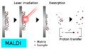 MALDI.png 508 × 291; 27 КБ
-
 Mass selective instability.gif 1801 × 2485; 206 КБ
-
 Mass Spec Sample Phase.png 733 × 598; 61 КБ
-
 MASS SPECTROMETER AND CHEVROLET ENGINE - NARA - 17472412.jpg 6214 × 4835; 24 МБ
-
 MASS SPECTROMETER AND CHEVROLET ENGINE - NARA - 17472440.jpg 4853 × 6232; 25,76 МБ
-
 Mass Spectrometer at NIH (24549823931).jpg 600 × 475; 49 КБ
-
 Mass Spectrometric Immunoassay.png 576 × 792; 106 КБ
-
 Mass spectrometry above the Arctic Circle. Tromso. August'15.jpg 5728 × 4000; 3,58 МБ
-
 Mass Spectrometry Quantitative Proteomic Workflows.png 1056 × 704; 113 КБ
-
 Mass-spectrograph.jpg 1600 × 1200; 89 КБ
-
 Mass-spectrograph.svg 596 × 596; 68 КБ
-
 MasSpecPen.jpg 2331 × 2447; 2,45 МБ
-
 Mcp-no.png 2000 × 917; 184 КБ
-
 MIKES schematic.jpg 1280 × 720; 41 КБ
-
 Miniature Quadrupole Mass Spectrometer.png 371 × 291; 38 КБ
-
 MS-Ready relationships.png 1961 × 904; 82 КБ
-
 Myotoxin mass spectra.png 712 × 519; 42 КБ
-
 NanoSIMS50 instrument diagram.png 1109 × 841; 36 КБ
-
 NovichokDetectionByLC-HRMSMS.svg 1280 × 720; 28 КБ
-
 Orbitrap.png 1176 × 785; 101 КБ
-
 Parfume ESTASI.tif 856 × 633; 1,58 МБ
-
 Paultrap.jpg 1027 × 827; 40 КБ
-
 Penning ionization.jpg 1097 × 411; 35 КБ
-
 Peptide mass fig.jpg 672 × 664; 154 КБ
-
 Photoionization Mechanism.png 681 × 439; 18 КБ
-
 PhysRevLett.120.152501.pdf 1275 × 1650, 6 сторінок; 1,34 МБ
-
 Pressure in mass spectrometer.png 1186 × 480; 152 КБ
-
 PTR mass spectrometer with quadrupol mass analyzer, schematics.png 780 × 527; 47 КБ
-
 PTR-MS z analizatorem kwadrupolowym, schemat.png 806 × 520; 42 КБ
-
 PTR-TOF MS traces concentrations diagram.jpg 562 × 274; 122 КБ
-
-
 QQQ scan modes SIM MSMS.png 1500 × 971; 1,71 МБ
-
 Quadrupole Potential Surface.gif 405 × 468; 1,8 МБ
-
 Quadrupole.svg 408 × 171; 30 КБ
-
 Quadrupolo.png 1292 × 543; 135 КБ
-
 Resolution in mass spectrometry.png 616 × 293; 2 КБ
-
 SALDI.jpg 960 × 720; 38 КБ
-
 Schema a blocchi MS.png 819 × 460; 8 КБ
-
 Schematic depiction of LC-MS MS procedure..jpg 661 × 600; 76 КБ
-

-
 Schematic illustration of SALDI instrument.JPG 960 × 720; 43 КБ
-
 Schematic of the thermospray probe and ion source.png 1177 × 575; 154 КБ
-
 Secteur magnétique.png 1520 × 658; 151 КБ
-
 Sercon 20-22 IRMS.jpg 3609 × 2406; 752 КБ
-
 Sheath Flow Interface.jpg 720 × 540; 172 КБ
-
 Sheathless Interface.jpg 720 × 540; 38 КБ
-
 Shimadzu Mixed annotated.jpg 4023 × 3447; 2,13 МБ
-
 Shimadzu Mixed.jpg 4023 × 3447; 2,02 МБ
-
 SILAC LC Peaks Scheme.png 983 × 718; 56 КБ
-
 Some miniature mass spectrometer systems.png 776 × 422; 43 КБ
-
 Spectrometrist in lab.svg 916 × 635; 217 КБ
-
 SPIDER-SequenceTagHomologySearchTool.JPG 482 × 183; 18 КБ
-
 Static secondary-ion mass spectrometry.gif 522 × 595; 9 КБ
-
 STATIC.SIMS.RICHA.2.GIF 896 × 313; 13 КБ
-
 STATIC.SIMS.RICHA.3.GIF 776 × 338; 10 КБ
-
 STATIC.SIMS.RICHA.5.GIF 632 × 372; 4 КБ
-
 Surface enhanced laser desorption ionization.png 1075 × 1158; 376 КБ
-
 Tandem Mass Spec Schematic Diagram.png 10 793 × 6046; 1,77 МБ
-
 Targeted mass spectrometry.gif 768 × 161; 11 КБ
-

-
 The Miniature Paul Ion Trap and board-level RF electronics.png 298 × 190; 111 КБ
-
 Thermo - Finnigan LCQ Mass Spectrometer (15797493459).jpg 638 × 479; 36 КБ
-
 Thermo - Finnigan LCQ Mass Spectrometer 2 (15797771267).jpg 638 × 479; 86 КБ
-
 Thermo - Finnigan LCQ Mass Spectrometer 3 (15797493769).jpg 638 × 479; 68 КБ
-
 Thermo - Finnigan LCQ Mass Spectrometer 4 (15983496555).jpg 638 × 479; 118 КБ
-
 Théorie du "Turn around time".jpg 448 × 505; 16 КБ
-
 Top-down vs bottom-up proteomics image.tif 1436 × 2495; 1,19 МБ
-
 Toxicology Research at FDA (NCTR 1412) (6009045826).jpg 3857 × 2562; 5,7 МБ
-
 Triplo quadrupolo.svg 1292 × 543; 62 КБ
-
 U.S. Department of Energy - Science - 395 010 003 (9474983458).jpg 1024 × 680; 91 КБ
-
 U.S. Department of Energy - Science - 395 014 001 (10293874364).jpg 2624 × 3179; 768 КБ
-
 U.S. Department of Energy - Science - 395 023 001 (9786528932).jpg 479 × 640; 48 КБ
-
 U.S. Department of Energy - Science - 395 024 002 (9404534462).jpg 640 × 480; 29 КБ
-
 U.S. Department of Energy - Science - 395 025 002 (9404534490).jpg 640 × 480; 27 КБ
-
 U.S. Department of Energy - Science - 395 037 001 (9508994875).jpg 833 × 554; 189 КБ
-
 Whole body by Mass Spectrometry Imaging.jpg 1089 × 464; 106 КБ
-
 XCMS alignment.png 705 × 544; 155 КБ
-
 ZooMS Publication Timeline.jpg 17 121 × 7476; 4,61 МБ
-
 ZooMS Schematic Diagram.jpg 3783 × 2756; 483 КБ
-
 CEC Model 21-101 mass spectrometer PP2008.038.004.jpg 750 × 605; 335 КБ
-
 CEC-103 Mass Spectrometer parts PP2005.007.009 detail.jpg 193 × 290; 24 КБ
-
 CEC-103 Mass Spectrometer parts PP2005.007.009.jpg 750 × 747; 272 КБ
-
 Sibyl Rock Computing Manual 1946.jpg 2625 × 3325; 4,83 МБ
-
 Sibyl Rock to Harold Wiley 1943.jpg 2550 × 3288; 6,47 МБ
-
 Пики с массой 28 Да при разных R.png 1157 × 353; 30 КБ
-
 Չհագեցվածության կանոն.png 1125 × 265; 28 КБ


_(9806908675).jpg)
.png)


_6486_(8755007149).jpg)
_6487_(8755005115).jpg)
_6489_(8755003029).jpg)
_6492_(8756122836).jpg)
_6496_(8756119526).jpg)
_6498_(8756116078).jpg)
_spectrometrist.svg)


.jpg)


.jpg)
.jpg)
.jpg)
.jpg)

_(6009045826).jpg)

.jpg)
.jpg)
.jpg)
.jpg)
.jpg)
.jpg)


_Mass_spectrometer.jpg)