Category:Microbiology
Przejdź do nawigacji
Przejdź do wyszukiwania
| Category Microbiology on sister projects: | |||||||||
|---|---|---|---|---|---|---|---|---|---|
Wiktionary |
Wikiversity |
Commons | |||||||
English: Microbiology is the study of microorganisms, today, work is largely done within the disciplines of biochemistry and genetics.
nauka o mikroorganizmach  | |||||
| Prześlij plik multimedialny | |||||
| Jest to | |||||
|---|---|---|---|---|---|
| Podklasa dla |
| ||||
| Składa się z | |||||
| |||||
Podkategorie
Poniżej wyświetlono 65 spośród wszystkich 65 podkategorii tej kategorii.
*
A
- Air microbiology (3 pliki)
- American Society for Microbiology (4 pliki)
B
C
D
E
- Endospore (1 strona, 16 plików)
- Eosin methylene blue (3 pliki)
G
- Gene Transfer Agents (8 plików)
H
- Hydrocarbonoclastic bacteria (1 plik)
I
- Industrial microbiology (14 plików)
L
M
- MELiSSA (2 pliki)
- Microbial art (5 plików)
- Microbial interactions (20 plików)
- Microbial metabolism (35 plików)
- Microbial toxins (5 plików)
- Microbial viability (31 plików)
N
P
- Plate Count Agar (3 pliki)
S
- Sabouraud Dextrose Agar (6 plików)
- Serotype (1 plik)
- Subviral particles (8 plików)
T
- Trophozoites (21 plików)
V
W
Pliki w kategorii „Microbiology”
Poniżej wyświetlono 200 spośród wszystkich 621 plików w tej kategorii.
(poprzednia strona) (następna strona)-
 12864 2008 Article 1790 Fig1 HTML.png 1184 × 486; 314 KB
-
 15-0339.R1-Cover.tif 1496 × 1936; 8,31 MB
-
 2 trophozoite of Gregarina Garnhami.jpg 1044 × 772; 100 KB
-
 2 trophozoites Gregarina Garnhami.jpg 1044 × 772; 108 KB
-
 2 trophozoites2.jpg 1044 × 772; 78 KB
-
 2016-07-01-sampling-Day 0-ESA S.Sechi-027 .jpg 1600 × 1200; 343 KB
-
 41598 2018 35246 Fig1 HTML.png 900 × 660; 294 KB
-
 500 Year Microbiology Experiment.JPG 2592 × 1936; 1,6 MB
-
 A Microbiologist.jpg 1439 × 2559; 188 KB
-
 A&B microbiological1.jpg 1160 × 300; 197 KB
-
 Acarbose degradation in gut microbiome.jpg 638 × 384; 68 KB
-
 Acid fast bacilli in sputum smear.jpg 1173 × 619; 663 KB
-
 Acid Product by Pneumonia 1.tif 1280 × 1024; 2,81 MB
-
 Acid Product by Pneumonia 2.tif 1280 × 1024; 3,03 MB
-
 Acid Product by Pneumonia 3.tif 1280 × 1024; 3,02 MB
-
 Adenovirus infection.jpg 1377 × 775; 137 KB
-
 Aeromonas veronii biovar sobria Gram Stain on Microscope Slide.jpg 1796 × 1796; 364 KB
-
 AFLP-patronen.jpg 844 × 415; 35 KB
-
 Agar Diffusion Method 1.jpg 961 × 514; 103 KB
-
 Agar Diffusion Method 2.jpg 960 × 720; 85 KB
-
 Agar en tub inclinat Kligler (KIA).jpg 1536 × 2048; 216 KB
-
 Agar XLD per la identificació de Salmonella.jpg 768 × 1024; 73 KB
-
 Aglutinació en porta.png 572 × 235; 58 KB
-
 Air sample room. Method of sedimentation. after 20 hours.png 3584 × 2016; 8,64 MB
-
 Air sample room. Method of sedimentation. after 36 hours.png 3584 × 2016; 8,25 MB
-
 Air sample room. Method of sedimentation. after 56 hours.png 3584 × 2016; 7,42 MB
-
 Air sample room. Method of sedimentation. after 66 hours.png 3584 × 2016; 7,2 MB
-
 Airborne contaminant.jpg 3202 × 1931; 2,27 MB
-
 Albert Jan Kluyver, his life and work (1959) (17760692188).jpg 1652 × 2852; 425 KB
-
 Alteranira Alternata.jpg 588 × 1280; 50 KB
-
 AMG 3724.jpg 3872 × 2581; 6,49 MB
-
 Annales de l'Institut Pasteur (1889) (18016503508).jpg 2816 × 1748; 1,98 MB
-
 Annales de l'Institut Pasteur (1889) (18177881536).jpg 3014 × 1744; 1,43 MB
-
 Annales de l'Institut Pasteur (1891) (17583765093).jpg 1992 × 3082; 961 KB
-
 Annales de l'Institut Pasteur (1891) (18016500808).jpg 1850 × 3138; 716 KB
-
 Annales de l'Institut Pasteur (1892) (17582084284).jpg 1110 × 2918; 427 KB
-
 Annales de l'Institut Pasteur (1892) (17584106053).jpg 2630 × 2205; 834 KB
-
 Annales de l'Institut Pasteur (1892) (18204715385).jpg 2840 × 2108; 890 KB
-
 Annales de l'Institut Pasteur (1894) (17582152394).jpg 1742 × 2834; 832 KB
-
 Annales de l'Institut Pasteur (1896) (17581587714).jpg 1664 × 2570; 801 KB
-
 Annales de l'Institut Pasteur (1896) (18016320318).jpg 1858 × 2968; 646 KB
-
 Annales de l'Institut Pasteur (1896) (18016323928).jpg 1990 × 2622; 1021 KB
-
 Annales de l'Institut Pasteur (1897) (17581826274).jpg 1824 × 2960; 1,12 MB
-
 Annales de l'Institut Pasteur (1897) (18200851312).jpg 1790 × 2834; 855 KB
-
 Annales de l'Institut Pasteur (1899) (14764609881).jpg 1940 × 2840; 1004 KB
-
 Annales de l'Institut Pasteur (1901) (18018342919).jpg 1872 × 2556; 877 KB
-
 Annales de l'Institut Pasteur (1902) (18016956090).jpg 1428 × 2860; 868 KB
-
 Annales de l'Institut Pasteur (1902) (18205812631).jpg 1268 × 2666; 412 KB
-
 Annales de l'Institut Pasteur (1903) (17581984004).jpg 1870 × 2830; 950 KB
-
 Annales de l'Institut Pasteur (1910) (18016891640).jpg 2638 × 1600; 995 KB
-
 Annales de l'Institut Pasteur (1910) (18017138200).jpg 1598 × 2878; 1,01 MB
-
 Annales de l'Institut Pasteur (1910) (18178239356).jpg 2736 × 704; 528 KB
-
 Annales de l'Institut Pasteur (1910) (18201248602).jpg 1592 × 2902; 1,67 MB
-
 Annales de l'Institut Pasteur (1910) (18205919611).jpg 2704 × 986; 789 KB
-
 Annales de l'Institut Pasteur (1912) (17582279884).jpg 1746 × 2848; 625 KB
-
 Annales de l'Institut Pasteur (1912) (17584118783).jpg 842 × 2540; 602 KB
-
 Annales de l'Institut Pasteur (1912) (17584197963).jpg 1888 × 2888; 1,27 MB
-
 Annales de l'Institut Pasteur (1912) (17584201723).jpg 952 × 2880; 743 KB
-
 Annales de l'Institut Pasteur (1912) (18016906748).jpg 1346 × 2704; 950 KB
-
 Annales de l'Institut Pasteur (1912) (18178410296).jpg 1816 × 2886; 666 KB
-
 Annales de l'Institut Pasteur (1912) (18201138202).jpg 1344 × 2706; 876 KB
-
 Annales de l'Institut Pasteur (1912) (18204650895).jpg 2752 × 1564; 1,12 MB
-
 Annales de l'Institut Pasteur (1912) (18204824685).jpg 1910 × 2850; 903 KB
-
 Annales de l'Institut Pasteur (1914) (17584188653).jpg 2992 × 1926; 1,55 MB
-
 Annales de l'Institut Pasteur (1914) (18017080860).jpg 2864 × 1750; 547 KB
-
 Annales de l'Institut Pasteur (1914) (18018418749).jpg 1442 × 2614; 502 KB
-
 Annales de l'Institut Pasteur (1914) (18018430889).jpg 1566 × 2650; 399 KB
-
 Annales de l'Institut Pasteur (1914) (18178334196).jpg 1366 × 2782; 829 KB
-
 Annales de l'Institut Pasteur (1914) (18204811055).jpg 1722 × 2768; 762 KB
-
 Annales de l'Institut Pasteur (1914) (18205827551).jpg 1580 × 2634; 372 KB
-
 Annales de l'Institut Pasteur (1914) (18205859241).jpg 2656 × 1130; 381 KB
-
 Annales de l'Institut Pasteur (1914) (18205987561).jpg 2704 × 1864; 1,19 MB
-
 Annales de l'Institut Pasteur (1918) (18016794760).jpg 2544 × 1672; 1 MB
-
 Annales de l'Institut Pasteur (1918) (18016953680).jpg 2002 × 3000; 929 KB
-
 Annales de l'Institut Pasteur (1918) (18178111166).jpg 1710 × 2552; 687 KB
-
 Annales de l'Institut Pasteur (1919) (14589838420).jpg 1382 × 1402; 442 KB
-
 Annales de l'Institut Pasteur (1919) (14589902208).jpg 1070 × 1454; 473 KB
-
 Annales de l'Institut Pasteur (1919) (14774159634).jpg 2976 × 1784; 841 KB
-
 Annales de l'Institut Pasteur (1919) (14796355873).jpg 2816 × 1576; 1,68 MB
-
 Annales de l'Institut Pasteur (1919) (14796412573).jpg 1084 × 1446; 513 KB
-
 Annales de microbiologie (1915) (17582307594).jpg 2688 × 1798; 1,12 MB
-
 Antimicrobial-resistant fungi.png 1107 × 815; 122 KB
-
 Antistof.JPG 255 × 360; 44 KB
-
 Antonio e Biagio e Cesare Arrigo Laboratory.jpg 3264 × 2448; 4,03 MB
-

-
-
-

-

-

-
-
-

-

-

-

-

-
 Ascitic fluid analysis-Findings.jpg 3264 × 2448; 1,95 MB
-
 ASLO.JPG 4288 × 3216; 4,08 MB
-
 ASO Slide.jpg 2048 × 1536; 367 KB
-
 Atalante Tahiti.jpg 500 × 375; 37 KB
-
 Autonomous Pathogen Detection System.jpg 256 × 384; 27 KB
-
 Auxanogramme API 20NE 2 PFraperie.pdf 800 × 518; 2,42 MB
-
 Auxanogramme FB.jpg 2048 × 2008; 225 KB
-

-

-
 Azo pic.tif 720 × 960; 891 KB
-
 B. subtilis sporulation reg.png 759 × 436; 131 KB
-
 Bacillus mycoides.jpg 4000 × 6000; 20,29 MB
-
 Bacteria gram negativa, Escherichia coli.jpg 1200 × 1600; 310 KB
-
 Bacteria with capsule.jpg 762 × 414; 170 KB
-
 Bacterial cell structure.png 802 × 399; 134 KB
-
 Bacterial colony.jpg 1535 × 2056; 192 KB
-
 Bacterial isolate.jpg 653 × 410; 16 KB
-
 Bacteriecel.png 175 × 287; 1 KB
-
 BD BACTEC FX40.jpg 4160 × 2340; 2,22 MB
-
 Bec-bunsen.png 356 × 435; 3 KB
-
 Beebe Biology.jpg 4608 × 2112; 604 KB
-
 Beebe MicrobeRocks.jpg 4608 × 2112; 729 KB
-
 Bench-scale Bioreactor.JPG 960 × 1280; 315 KB
-
 Bilal D.jpg 2576 × 1932; 1,73 MB
-
 Bilal Djeghout Laboratory.jpg 1966 × 2048; 361 KB
-
 Bile esculin agar test.jpg 1552 × 2300; 1,67 MB
-
 Bimssteine4056.jpg 531 × 384; 48 KB
-
 Biodegradation of Pollutants.png 1524 × 862; 178 KB
-
 Bioréacteur CiYOU.png 1883 × 952; 2,9 MB
-
 Blood and chocolate agar piles.jpg 4160 × 2340; 2,25 MB
-
 Blood corpuscles. Wellcome L0006532.jpg 1106 × 1718; 1,07 MB
-
 Blood corpuscles. Wellcome L0006533.jpg 1090 × 1726; 938 KB
-
 BOD Incubator.jpg 1844 × 4000; 3,77 MB
-
 Brettanomyces colonies.JPG 3648 × 2736; 3,73 MB
-
 Brettanomyces plate 01.jpg 2348 × 1664; 1,25 MB
-
 Brettanomyces plate 02.JPG 3648 × 2736; 3,84 MB
-
 Brettanomyces plate 03.JPG 3648 × 2736; 3,77 MB
-
 Brettanomyces plate 04.JPG 3648 × 2736; 3,7 MB
-
 Brettanomyces plate 05.JPG 3648 × 2736; 3,71 MB
-
 Brettanomyces plate 06.JPG 3648 × 2736; 3,73 MB
-
 Brettanomyces plate 07.JPG 3648 × 2736; 3,81 MB
-
 Brettanomyces plate 08.JPG 3648 × 2736; 3,73 MB
-
 Brettanomyces plate 09.JPG 3648 × 2736; 3,82 MB
-
 Brettanomyces plate 10.JPG 3648 × 2736; 3,78 MB
-
 Brettanomyces plate 11.JPG 3648 × 2736; 3,75 MB
-
 Brettanomyces plate 12.jpg 3392 × 2236; 2,62 MB
-
 Brettanomyces plate 13.jpg 3648 × 2380; 2,56 MB
-
 Brettanomyces plate 14.JPG 3648 × 2736; 3,73 MB
-
 Brettanomyces plate 15.jpg 3648 × 2608; 2,68 MB
-
 Bronze inkstand used by Louis Pasteur, Europe, 1801-1830 Wellcome L0057303.jpg 4256 × 2832; 1,98 MB
-
 BSc in Lab Medicine Part-I.pdf 1275 × 2100, 79 stron; 1,22 MB
-
 BSC line.jpg 1210 × 1806; 513 KB
-
 BSC with microscope.jpg 1960 × 1490; 940 KB
-

-
 Bunte Reihe Microplate.jpg 1510 × 1078; 458 KB
-
 C.perfringens.png 750 × 752; 817 KB
-
 Camara Neubauer.jpg 400 × 267; 11 KB
-
 Candida albicans AO AL.jpg 1280 × 960; 565 KB
-
 Candida albicans growing as yeast cells and filamentous (hypha) cells.jpg 5152 × 3864; 5,01 MB
-
 Capnocytophaga canimorsus on blood agar 2.jpg 1744 × 1746; 455 KB
-
 Carbohydrate fermentation gonorrhea.png 314 × 296; 28 KB
-
 Carbon biogeochemical cycle.png 896 × 574; 156 KB
-
 Caulimovirus Life Cycle.png 448 × 410; 19 KB
-
 CAVES-Microbiology-Day0-S.Sechi-1060178 .jpg 1600 × 1067; 264 KB
-
 Chamberland filter, France, 1875-1884 Wellcome L0057279.jpg 4288 × 2848; 976 KB
-
 Cheese rind microbial community.png 2133 × 1600; 186 KB
-
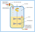 Chemostat Vessel Diagram.png 543 × 497; 34 KB
-
 Chlamydospore of Candida albicans in LPCB preparation.jpg 4160 × 2340; 1,72 MB
-

-

-

-
 Chlorella with yellow indicative of chlorophyll.jpg 396 × 400; 14 KB
-
 Chtx-Deu4.png 960 × 720; 35 KB
-
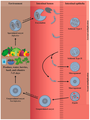 Cicle vital de Cyclospora Cayetanensis.png 2730 × 3627; 2,26 MB
-
 Ciclo.JPG 431 × 206; 11 KB
-
 Citric Acid Cycle Illustration.jpg 2934 × 2197; 1,11 MB
-
 Cluster of PAOs.jpg 4080 × 3072; 3,18 MB
-
 Codonosiga botrytis.jpg 202 × 334; 37 KB
-
 Coleção de Culturas de Fitobactérias - APTA - SAASP.jpg 640 × 480; 154 KB
-
 Colonia fungina 01.jpg 1555 × 1166; 812 KB
-
 Colonia fungina 02.jpg 1077 × 808; 487 KB
-
 Colorimeter -Micrbiology depertment instrument bankura sammilani college.jpg 1920 × 2560; 332 KB
-
 Complement pathway.png 688 × 834; 89 KB
-
 Composición de la PM V.png 217 × 111; 42 KB
-

-
-
 Conjugacion bacteriana.jpg 490 × 261; 40 KB
-
 Conjugatie (genetica).png 2708 × 1434; 1,26 MB
-
 Construct.jpg 2400 × 3000; 298 KB
-
 Continuous Culture System.jpg 2400 × 2400; 221 KB
-
 Control after 72 h.tif 1440 × 1024; 4,25 MB
-
 Controllo Salmonella.png 692 × 1032; 100 KB
-
 Cool Science Glasses.jpg 480 × 640; 80 KB
-
-

-
 Corneal scraping with fungal elements.jpg 1500 × 804; 519 KB
-
 Corynebacterium diphtheriae albert stain.jpg 2592 × 1456; 963 KB
-
 Corynebacterium Gram.JPG 4288 × 3216; 3,06 MB
-
 CourbeEtalonPCRQuanti.JPG 1276 × 1120; 757 KB
-
 CourbesFusionLeiden.JPG 1467 × 1105; 776 KB
-
 CoverSlips.jpg 4007 × 2671; 2,16 MB
-
 Crescita batterica su agar.jpg 3198 × 4799; 9,26 MB
-
 Critical Reviews in Microbiology.jpg 150 × 197; 21 KB


.jpg)


_(17760692188).jpg)


_(18016503508).jpg)
_(18177881536).jpg)
_(17583765093).jpg)
_(18016500808).jpg)
_(17584106053).jpg)
_(18204715385).jpg)
_(17582152394).jpg)
_(17581587714).jpg)
_(18016320318).jpg)
_(18016323928).jpg)
_(17581826274).jpg)
_(18200851312).jpg)
_(14764609881).jpg)
_(18018342919).jpg)
_(18016956090).jpg)
_(18205812631).jpg)
_(17581984004).jpg)
_(18016891640).jpg)
_(18017138200).jpg)
_(18201248602).jpg)
_(17582279884).jpg)
_(17584197963).jpg)
_(18016906748).jpg)
_(18178410296).jpg)
_(18201138202).jpg)
_(18204650895).jpg)
_(18204824685).jpg)
_(17584188653).jpg)
_(18017080860).jpg)
_(18018418749).jpg)
_(18018430889).jpg)
_(18178334196).jpg)
_(18204811055).jpg)
_(18205827551).jpg)
_(18205859241).jpg)
_(18205987561).jpg)
_(18016794760).jpg)
_(18016953680).jpg)
_(18178111166).jpg)
_(14589838420).jpg)
_(14589902208).jpg)
_(14774159634).jpg)
_(14796355873).jpg)
_(14796412573).jpg)
_(17582307594).jpg)


_(19122191244).jpg)
_(19122209334).jpg)
_(19122223144).jpg)
_(19123893243).jpg)
_(19123899183).jpg)
_(19556705168).jpg)
_(19556716868).jpg)
_(19718566316).jpg)
_(19718593096).jpg)
_(19718596636).jpg)
_(19744816495).jpg)
_(19749441651).jpg)


_cells.jpg)


_(ABCDEGHJ).png)
_(E)_thorne_aka_spike.png)
.png)


.png)


_(17582084284).jpg)
_(18178239356).jpg)
_(18205919611).jpg)
_(17584118783).jpg)
_(17584201723).jpg)
,encoding_the_large_clostridial_toxins(LCTs)_involved_in_C._difficile_infections_CDI.png)