Category:Parasites
Jump to navigation
Jump to search

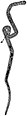
organism adapted to living on or in another organism and causing harm to its host _parasitized_by_Acrodactyla_quadrisculpta_larva_(RMNH.INS.593867)_-_BDJ.1.e992.jpg) | |||||
| Upload media | |||||
| Subclass of | |||||
|---|---|---|---|---|---|
| Part of | |||||
| Named after |
| ||||
| Different from | |||||
| |||||
Subcategories
This category has the following 62 subcategories, out of 62 total.
- Parasite evolution (35 F)
- Parasite physiology (46 F)
*
+
- Media from Parasites & Vectors (51 F)
.
A
B
C
D
E
F
- Foleyella furcata (3 F)
G
- Gyrodactylus salaris (4 F)
H
M
- Meguro Parasitological Museum (36 F)
- Merozoites (30 F)
N
O
- Oeciacus hirundinis (2 F)
P
- Parasitic protozoans (38 F)
- Placobdelloides siamensis (13 F)
- Pterygodermatites quentini (6 F)
R
- Rhynchophorus vulneratus (7 F)
- Ricinus vaderi (4 F)
S
- Sporozoites (19 F)
- Stomoxys calcitrans (51 F)
T
- Tunga caecata (2 F)
V
Pages in category "Parasites"
The following 5 pages are in this category, out of 5 total.
Media in category "Parasites"
The following 200 files are in this category, out of 293 total.
(previous page) (next page)-
 13071 2019 3727 Fig1.webp 1,772 × 1,097; 229 KB
-
 2020-04-07 - Tick 01.jpg 880 × 1,408; 113 KB
-
 A pure parasite.jpg 2,592 × 1,556; 944 KB
-
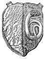 A worm found in the left ventricle Wellcome L0012373.jpg 1,212 × 1,582; 856 KB
-
 Actinomyxon zh hans.png 644 × 1,092; 9 KB
-
 Alpenrosen-Apfel.jpg 1,024 × 609; 85 KB
-
 Ameisensackkaefer mit Wespen bei Eiablage crop 2020.jpg 4,000 × 2,667; 5.4 MB
-
 Antistrophus jeanae larvae eating inside Silphium perfoliatum.jpg 2,222 × 1,336; 606 KB
-
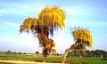
 Arbre dans la ville de Tchamba 2.jpg 2,160 × 3,840; 3.61 MB
-
 Arbre dans la ville de Tchamba 3.jpg 2,160 × 3,840; 3.43 MB
-
 Arbre dans la ville de Tchamba 4.jpg 2,160 × 3,840; 3.25 MB
-
 Arbre dans la ville de Tchamba.jpg 2,160 × 3,840; 3.17 MB
-
 Archives slaves de biologie BHL31440927.jpg 2,247 × 3,528; 601 KB
-
 Bed bug dead.jpg 1,656 × 1,281; 520 KB
-
 Bestioles non identifiables 05.jpg 2,112 × 2,112; 733 KB
-
 Bestioles non identifiables 06.jpg 2,112 × 2,112; 811 KB
-
 Bestioles non identifiables 07.jpg 2,112 × 2,112; 737 KB
-
 Bestioles non identifiables 10.jpg 2,112 × 2,112; 944 KB
-
 Bestioles non identifiables 11.jpg 2,112 × 2,112; 681 KB
-
 Bestioles non identifiables 16.jpg 2,112 × 2,112; 834 KB
-
 Bestioles non identifiables 17.jpg 2,112 × 2,112; 747 KB
-
 Bestioles non identifiables 18.jpg 2,112 × 2,112; 822 KB
-
 Bilhazia parasite, Manson Wellcome M0012602.jpg 2,650 × 4,258; 2.29 MB
-
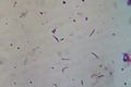
 Blood-smear-showing-p-relictum-infection-stained-purple-within-red-blood-cells.jpg 3,072 × 2,048; 208 KB
-
 Bronze inkstand used by Louis Pasteur, Europe, 1801-1830 Wellcome L0057303.jpg 4,256 × 2,832; 1.98 MB
-
 Calydiscoides euzeti.jpg 1,199 × 3,455; 980 KB
-
 CAmericanaMar03.jpg 858 × 1,200; 211 KB
-
 Camponotus morosus ant being attacked by a parasitoid phorid fly.jpg 358 × 239; 21 KB
-
 Capillaria alcoveri male Figure 10.png 1,278 × 1,735; 41 KB
-
 Capillaria bainae Justine & Radujkovic 1988 Figure 2G & 2H.png 786 × 961; 27 KB
-
 Capillaria bainae Justine & Radujkovic 1988 Figure 2G.png 450 × 961; 17 KB
-
 Capillaria bainae Justine & Radujkovic 1988 Figure 2H.png 404 × 753; 10 KB
-
 Capillaria brochieri Justine, 1988 - Male extremity.png 1,012 × 972; 26 KB
-
 Capillaria legerae male Figure 6B.png 790 × 1,196; 33 KB
-
 Capillaria myoxinitelae female Figure 2F.png 628 × 784; 20 KB
-
 Capillaria myoxinitelae male Figure 2B.png 740 × 760; 13 KB
-
 Carnus hemapterus IB.jpg 480 × 307; 27 KB
-
 Carnus hemapterus.JPG 2,091 × 1,429; 174 KB
-
 Cephalothorax of a female R. Teagueselfi.png 412 × 535; 239 KB
-
 Children outside straw hut Wellcome L0040944.jpg 3,899 × 2,630; 6.11 MB
-
 Chrysomya-bezziana-adults-myiasis-larvae-2.jpg 800 × 483; 126 KB
-
 Clamps of the posterior end of Sparicotyle chrysophrii.tif 2,272 × 1,704; 11.08 MB
-
 CochenillesJuinTilleul2010 2.jpg 2,180 × 1,556; 536 KB
-
 CochenillesJuinTilleul2010 3.jpg 2,128 × 1,328; 445 KB
-
 CochenillesJuinTilleul2010.jpg 737 × 481; 83 KB
-
 Coracidium.PNG 570 × 460; 161 KB
-
 Cordyceps - Lilly Pilly Gully, Wilson's Promotory National Park.jpg 3,024 × 4,032; 2.39 MB
-
Crab louse 01.webm 19 s, 320 × 240; 1.18 MB
-
Crab louse 02.webm 39 s, 320 × 240; 1.11 MB
-
 Cuscuta europaea (on Urtica dioica).jpg 2,649 × 3,531; 2.13 MB
-
 Cyst of Entamoeba histolytica at a magnification of 1600X.jpg 3,264 × 2,448; 960 KB
-
 Delimitation of subspecies of bats by DNA barcoding of their ectoparasites.jpg 2,128 × 1,050; 407 KB
-
 Dictyostega Flora brasiliensis Vol III Part I Fasc 8 Plate 7 Fig IV.jpg 685 × 574; 112 KB
-
 Dracunculus medinensis larvae.jpg 700 × 474; 51 KB
-
 Dryiniden Säckchen.png 408 × 552; 29 KB
-
 Dujardin 1845 Planche 2.png 2,604 × 4,385; 12.04 MB
-
 E. May, "Relation ...monster or serpent", text Wellcome L0012375.jpg 1,036 × 1,817; 583 KB
-
 E. May, "Relation ...monster or serpent", text Wellcome L0012377.jpg 1,038 × 1,834; 591 KB
-
 E. May, "Relation ...monster or serpent", text Wellcome L0012378.jpg 1,002 × 1,932; 1.01 MB
-
 E. May, "Relation ...monster or serpent", text Wellcome L0012379.jpg 974 × 1,802; 940 KB
-
 EB1911 Gregarines - Porospora gigantea.jpg 251 × 1,083; 58 KB
-

-
 Echinococcus multilocularis.jpg 1,258 × 817; 25 KB
-
 Eggs of Sparicotyle chrysophrii.tif 2,272 × 1,704; 11.08 MB
-

-
 Entamoeba histolytica life cycle-en.svg 1,029 × 976; 267 KB
-
 Enterobius vermicularis Life Cycle.png 1,171 × 815; 155 KB
-
 Entoloma.abortivum.001.jpg 960 × 720; 70 KB
-
 Euphrasia officinalis at Spier's.jpg 1,712 × 2,288; 1.34 MB
-
 Euplectrus sp. - lifecycle A - 06 - imagines (2009-05-10).jpg 1,700 × 1,181; 175 KB
-
 European Mistletoe Growing On Trees.jpg 1,280 × 853; 615 KB
-
 Extpost macho a galli 1.jpg 976 × 733; 166 KB
-
 Extpost macho a galli 2.jpg 983 × 731; 168 KB
-
 Fascioliasis lifecycle.png 578 × 564; 235 KB
-
-
 Fmars-08-720424-g001.jpg 2,285 × 1,545; 219 KB
-

-

-

-
 Gardia lamblia cyst.JPG 745 × 513; 64 KB
-
 Geographic MOSAIC MODEL of Coevolution.jpg 548 × 413; 128 KB
-
 Gigantocotyle formosanum.jpg 349 × 585; 148 KB
-
 H.nana livscykel, redigerad bild.jpg 520 × 435; 28 KB
-
 Haematopinus tuberculatus.tif 2,584 × 1,936; 8.97 MB
-
 Heteromicrocotyloides megaspinosus Body.jpg 1,772 × 2,843; 525 KB
-
 Heterorhabditis downesi.jpg 710 × 595; 100 KB
-
 Hexó.png 653 × 496; 65 KB
-
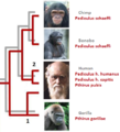 Hominine lice.png 408 × 450; 135 KB
-
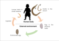
 Host parasite interaction.jpg 4,000 × 2,250; 1.15 MB
-
 Huffmanela branchialis.jpg 848 × 452; 79 KB
-
 Huffmanela filamentosa.jpg 1,376 × 633; 80 KB
-
 Huffmanela hamo egg - microscope line drawing.png 982 × 673; 7 KB
-
 Huffmanela hamo eggs in Muraenesox cinereus 1A.JPG 3,264 × 2,448; 1.63 MB
-
 Huffmanela hamo eggs in Muraenesox cinereus 1B.JPG 2,048 × 1,536; 870 KB
-
 Huffmanela hamo eggs in Muraenesox cinereus 1C.JPG 2,048 × 1,536; 910 KB
-
 Huffmanela lata.jpg 968 × 756; 125 KB
-
 Huffmanela ossicola.jpg 1,048 × 666; 145 KB
-
 Infection cycle of a Rhinophoridae fly in a woodlouse host.jpg 1,512 × 901; 392 KB
-
 Inquicus fellatus.jpg 322 × 578; 410 KB
-
 Issus.coleoptratus.nymph.with.Dryininae.larva.jpg 1,270 × 1,282; 184 KB
-
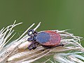 Ixodes ricinus on dry grass.jpg 2,155 × 1,616; 1.77 MB
-
 Ixodholfem5.jpg 320 × 451; 20 KB
-
 J. Cropper, A phenomenal abundance of parasi Wellcome L0032682.jpg 5,286 × 3,556; 4.93 MB
-
 Jmse-11-00001-g006+.jpg 1,628 × 2,496; 846 KB
-
 Jmse-11-00001-g006.webp 1,707 × 2,772; 2.36 MB
-
 Jmse-11-00001-g007+.jpg 1,723 × 2,357; 471 KB
-
 Jmse-11-00001-g007.webp 1,818 × 2,375; 1.1 MB
-
 Jmse-11-00001-g007a.jpg 1,725 × 1,204; 346 KB
-
 Kissing bug hut Wellcome L0040945.jpg 4,008 × 2,672; 5.01 MB
-
 Komodo8 1-1-12 - 10 wicked parasite (6695814383).jpg 1,134 × 853; 152 KB
-
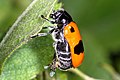
 Ladybug w larva.jpg 3,264 × 2,448; 2.51 MB
-
 Larvae of Strongyloides from culture.jpg 4,000 × 2,250; 919 KB
-
 Leshmania infantum.JPG 1,473 × 985; 144 KB
-
 Leucochloridium paradoxum metacercaria from Heckert 1889 plate1 fig5.png 541 × 795; 488 KB
-
 Leucochloridium paradoxum sporocyst from Heckert 1889 plate1 fig1.png 590 × 964; 634 KB
-
 Life cycle.JPG 689 × 514; 59 KB
-
 Life-cycle stages of the parasite Haemogregarina muris and i Wellcome V0022562.jpg 2,418 × 3,096; 3.48 MB
-
 Maconellicoccus hirsutus - hibiscus mealybug - adult male.jpg 616 × 634; 249 KB
-
 Male and female haem columbae.jpg 3,648 × 2,736; 2.09 MB
-
 Mallophaga Life Cycle.jpg 1,800 × 1,359; 381 KB
-
 Mare-de-rovello.jpg 1,200 × 1,600; 271 KB
-
 Mealybug on Phalaenopsis.jpg 1,200 × 1,076; 431 KB
-
 Mela&bee.tif 1,037 × 705; 2.1 MB
-
 MelaAttack.jpg 350 × 197; 62 KB
-
 Merlup1.JPG 2,848 × 2,136; 1.34 MB
-
 Micropredator Parasite Parasitoid Predator strategies compared.svg 1,857 × 648; 156 KB
-
 Microscopy; various parasites at 1000X magnification. Colour Wellcome V0024968.jpg 2,310 × 3,186; 3.55 MB
-
 Mid-posterior end of Sparicotyle chrysophrii.tif 2,272 × 1,704; 11.08 MB
-
 Monocystis Capture.jpg 602 × 627; 60 KB
-
 Moravecnema segonzaci.jpg 1,193 × 1,386; 233 KB
-
 Nature's umbrella (Mushrooms).jpg 2,592 × 1,728; 1.72 MB
-
 Negative Frequencie Dependant Selection.jpg 723 × 448; 41 KB
-

-
 Nerocila orbignyi k.jpg 320 × 240; 58 KB
-
 Ophiocordyceps unilateralis, Molecular and Chemical Functions.png 4,800 × 2,700; 6.36 MB
-
 Parasite (journal) 2013 cover 1000pix.jpg 707 × 1,000; 119 KB
-
 Parasite (journal) 2013, 20, 23 Veciana - Physaloptera.pdf 1,233 × 1,747, 5 pages; 1.38 MB
-
 Parasite (journal) 2013, 20, 32 Karadjian - Haemoproteus.pdf 1,233 × 1,747, 11 pages; 2.9 MB
-

-
-

-
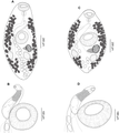
 Parasite 20,56 (2013) Neidhartia & Prosorhynchus figs 1-6 png600.png 4,088 × 6,680; 4.49 MB
-
 Parasite filled blood.jpg 2,688 × 1,520; 897 KB
-
 Parasite journal) 2013, 20, 43, Watermeyer, Setaria.pdf 1,233 × 1,747, 8 pages; 2.26 MB
-
 Parasite on Persea americana tree.jpg 2,990 × 4,138; 3.93 MB
-
 Parasite.JPG 2,592 × 1,944; 1.47 MB
-
 Parasite130113-fig1 Haemonchotolerance in West African Dwarf goats.png 447 × 615; 364 KB
-
 Parasite130113-fig2 Haemonchotolerance in West African Dwarf goats.png 473 × 612; 541 KB
-
 Parasite130113-fig3 Haemonchotolerance in West African Dwarf goats.tif 1,102 × 924; 199 KB
-
 Parasite130113-fig4 Haemonchotolerance in West African Dwarf goats.tif 1,102 × 921; 117 KB
-
 Parasite130113-fig5 Haemonchotolerance in West African Dwarf goats.tif 1,093 × 773; 109 KB
-
 Parasite130113-fig6 Haemonchotolerance in West African Dwarf goats.tif 1,102 × 888; 130 KB
-
 Parasite130113-fig7 Haemonchotolerance in West African Dwarf goats.tif 1,102 × 844; 115 KB
-
 Parasite130116-2-olm Entamoeba gingivalis.pdf 1,239 × 1,754, 5 pages; 47 KB
-
 Parasite130116-fig1 Entamoeba gingivalis.tif 2,067 × 2,086; 1.02 MB
-
 Parasite130116-fig2 Entamoeba gingivalis.tif 1,102 × 980; 125 KB
-
 Parasite130116-fig3 Entamoeba gingivalis.tif 941 × 1,919; 277 KB
-
 Parasite130116-fig4 Entamoeba gingivalis.tif 1,102 × 926; 116 KB
-
 Parasite130116-fig5 Entamoeba gingivalis.tif 1,103 × 916; 174 KB
-
 Parasite130116-fig6 Entamoeba gingivalis.tif 1,102 × 1,102; 95 KB
-

-

-
 Parasite140013-fig1 Pterygodermatites (Paucipectines) baiomydis drawings.tif 2,343 × 2,624; 967 KB
-
 Parasite140013-fig3 Pterygodermatites (Paucipectines) baiomydis Photo.tif 1,102 × 896; 1.68 MB
-
 Parasite140065-fig3 Tree of hemogregarines.tif 2,067 × 2,417; 1.16 MB
-

-

-

-
 Parasite140113-fig1 Pea crab Nepinnotheres novaezelandiae in mussel.png 2,677 × 1,182; 387 KB
-
 Parasite140113-fig2 Pea crab Nepinnotheres novaezelandiae in mussel.png 1,102 × 583; 71 KB
-
 Parasite140113-fig3 Pea crab Nepinnotheres novaezelandiae in mussel.png 1,890 × 906; 172 KB
-
 Parasite140113-fig4 Pea crab Nepinnotheres novaezelandiae in mussel.png 1,890 × 914; 487 KB
-
 Parasite140113-fig5 Pea crab Nepinnotheres novaezelandiae in mussel.png 1,102 × 713; 100 KB
-
 Parasite140113-fig6 Pea crab Nepinnotheres novaezelandiae in mussel.png 1,102 × 727; 82 KB
-
 Parasite140113-fig7 Pea crab Nepinnotheres novaezelandiae in mussel.png 1,649 × 1,131; 118 KB
-
 Parasite140131-fig1 Capillaria plectropomi Figure 1H Caudal end of male.png 1,243 × 1,210; 178 KB
-
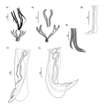
 Parasite140132-fig4 Philometra sp. (Nematoda, Philometridae) from Lutjanus johnii.png 2,067 × 1,718; 483 KB
-
 Parasite150050-fig1 genus Odilia (Nematoda, Heligmonellidae, Nippostrongylinae).tif 2,520 × 1,639; 168 KB
-
 Parasite150050-fig2 genus Odilia (Nematoda, Heligmonellidae, Nippostrongylinae).tif 2,362 × 2,312; 668 KB
-
 Parasite150050-fig3 genus Odilia (Nematoda, Heligmonellidae, Nippostrongylinae).tif 2,447 × 2,489; 800 KB
-
 Parasite150069-fig1 Aristocleidus mexicanus (Monogenea, Dactylogyridae).tif 1,654 × 1,987; 472 KB
-
 Parasite150069-fig2 Aristocleidus lacantuni (Monogenea, Dactylogyridae).tif 2,835 × 1,038; 205 KB
-
-
 Parasite180015-fig3 Sicuophora multigranularis (Armophorea, Clevelandellida).png 2,362 × 3,071; 2.05 MB
-
 Parasite180015-fig4 Sicuophora multigranularis (Armophorea, Clevelandellida).png 3,307 × 2,362; 2.87 MB
-
 Parasite180015-fig6 Tree of ciliates from the class Armophorea.png 3,543 × 2,676; 2.49 MB
-
 Parasite210036-fig2 - Creptotrema (Digenea, Allocreadiidae).png 4,692 × 5,275; 3.96 MB
-
 Parasite210036-fig4 - Creptotrema conconae (Digenea, Allocreadiidae).png 2,797 × 6,438; 3.64 MB
-
 Parasite210036-fig5 - Creptotrema schubarti (Digenea, Allocreadiidae).png 3,193 × 5,533; 3.47 MB
-
 Parasite210036-fig6 - Creptotrema megacetabularis (Digenea, Allocreadiidae).png 4,021 × 5,906; 3.81 MB
-
 Parasite210036-fig7 - Creptotrema (Digenea, Allocreadiidae).png 5,298 × 5,408; 2.74 MB
-
 Parasite210036-fig8 - Creptotrema (Digenea, Allocreadiidae).png 5,337 × 5,131; 1.38 MB
-
 Parasite210069-fig1 - Hassalstrongylus dollfusi (Nematoda, Heligmonellidae).tif 2,102 × 2,102; 1.16 MB
-
 Parasite210069-fig2 - Hassalstrongylus dollfusi (Nematoda, Heligmonellidae).png 8,400 × 8,400; 7.53 MB
-
 Parasite210069-fig3 - Hassalstrongylus dollfusi (Nematoda, Heligmonellidae).png 8,411 × 8,411; 6.28 MB
-
 Parasite210069-fig4 - Hassalstrongylus dollfusi (Nematoda, Heligmonellidae).png 8,410 × 8,411; 5.84 MB
-
 Parasite210069-fig6 - Hassalstrongylus dollfusi (Nematoda, Heligmonellidae).png 8,410 × 3,595; 1.98 MB
-
 Parasite210080-Fig01 Eutarsopolipus paryavae.png 3,972 × 5,339; 2.03 MB
-
 Parasite210080-Fig02 Eutarsopolipus paryavae.png 4,164 × 5,757; 2.21 MB
-
 Parasite210080-Fig03 Eutarsopolipus paryavae.png 4,329 × 5,240; 2.31 MB


.jpg)


.jpg)


_parasite_in_the_tentacles_of_Succines_putris_L.jpeg)


.jpg)


.jpg)


_2013_cover_1000pix.jpg)
_Neidhartia_%26_Prosorhynchus_figs_1-6_png600.png)


_from_Lutjanus_johnii.png)
,_parasites_of_cephalopods_of_the_Mediterranean_Sea.png)
.png)
.png)

.png)
.png)
.png)
.png)
.png)
.png)
.png)
.png)
.png)
.png)


