Category:Microscopic images of plants - cross sections of leaves
Jump to navigation
Jump to search
Subcategories
This category has the following 15 subcategories, out of 15 total.
A
B
Media in category "Microscopic images of plants - cross sections of leaves"
The following 108 files are in this category, out of 108 total.
-
Aerenchyma in hydrophyte leaf (33958474393).jpg 3,264 × 1,840; 1.31 MB
-
American journal of pharmacy (1883) (14771762412).jpg 3,856 × 2,392; 2.65 MB
-
Anatomia foliar.jpg 800 × 600; 83 KB
-
Angiosperm Leaf Abaxial Midrib Collenchyma in Nymphaea Leaf (36909776673).jpg 3,264 × 1,840; 952 KB
-
Angiosperm Leaf Adaxial Epidermis and Cuticle in Nerium (37814260526).jpg 3,264 × 1,840; 4.71 MB
-
Angiosperm Leaf Leaf Tip in Nerium (24010128358).jpg 3,264 × 1,840; 4.06 MB
-
Angiosperm Leaf Mesophyll Arrangement in Leaf Blade of Nymphea (37531059286).jpg 3,264 × 1,840; 1,004 KB
-
Angiosperm Leaf Mesophyll Arrangement in Nymphaea Midrib (23727157938).jpg 3,264 × 1,840; 1.2 MB
-
Angiosperm Leaf Mesophyll in Nerium (34321745784).jpg 3,264 × 1,840; 1.21 MB
-
Angiosperm Leaf Midrib Abaxial Collenchyma in Nerium (37862993931).jpg 3,264 × 1,840; 1.1 MB
-
Angiosperm Leaf Palisade Mesophyll in Nymphea (36909777763).jpg 3,264 × 1,840; 1.01 MB
-
Angiosperm Leaf Perivascular Tannin Cells in Nymphea Leaf (36909775853).jpg 3,264 × 1,840; 1.14 MB
-
Angiosperm Morphology Epidermis in the Xerophytic Leaf of Larrea (37431960475).jpg 3,264 × 1,840; 5.03 MB
-
Angiosperm Morphology Idioblasts in the Xerophytic Leaf of Larrea (36619801473).jpg 3,264 × 1,840; 1.7 MB
-
Angiosperm Morphology Lithocyst in Upper Epidermis in Ficus Leaf (35741364504).jpg 3,264 × 1,840; 4.05 MB
-
Angiosperm Morphology Lower Epidermis in Ficus Leaf (36376635982).jpg 3,264 × 1,840; 6.68 MB
-
Angiosperm Morphology Lower Epidermis in Ficus Leaf (36529923876).jpg 3,264 × 1,840; 4.22 MB
-
Angiosperm Morphology Mesophyll Arrangement in Ficus Leaf (35741363824).jpg 3,264 × 1,840; 5.79 MB
-
Angiosperm Morphology Spongy Mesophyll in Ficus Leaf (36376645382).jpg 3,264 × 1,840; 6.65 MB
-
Angiosperm Morphology Starch Sheath in the Xerophytic Leaf of Larrea (37033297610).jpg 3,264 × 1,840; 1.49 MB
-
Angiosperm Morphology Substomatal Chambers in Zea Leaf (37216538000).jpg 3,264 × 1,840; 1,004 KB
-
Angiosperm Morphology The Xerophytic Dicotyledonous Leaf of Ficus (35741366204).jpg 3,264 × 1,840; 6.49 MB
-
Angiosperm Morphology The Xerophytic Dicotyledonous Leaf of Ficus (36376641102).jpg 3,264 × 1,840; 5.89 MB
-
Angiosperm Morphology Upper Epidermis in Ficus Leaf (36376650822).jpg 3,264 × 1,840; 7.12 MB
-
Astrosklereide 140.jpg 600 × 414; 139 KB
-
Bifacial leaf cross section.jpg 678 × 800; 453 KB
-
Blattquerschnitt.jpg 1,280 × 1,024; 431 KB
-
Brockhaus and Efron Encyclopedic Dictionary b75 239-0.jpg 680 × 862; 93 KB
-
Cotton Leaf C.S. x50 magnification.png 739 × 663; 927 KB
-
Cotton Leaf C.S.png 733 × 721; 1.13 MB
-
Coupe transversale d'une feuille de gymnosperme.jpg 1,536 × 2,048; 469 KB
-
Crassula ovata , histology.JPG 1,024 × 770; 177 KB
-
Crassula ovata, histology.JPG 1,024 × 769; 285 KB
-
Cuticle overlying upper epidermis in mesophyte leaf (35103215772).jpg 3,264 × 1,840; 1.23 MB
-
Cuticle overlying upper epidermis of mesophyte leaf (34686901852).jpg 3,264 × 1,840; 1.03 MB
-
Cycas revoluta.tif 4,405 × 1,852; 19.58 MB
-
Cystolith in the leaf of Ficus Elastica.jpg 433 × 545; 126 KB
-
Die Gartenlaube (1892) b 798.jpg 1,569 × 609; 247 KB
-
Falcaria vulgaris sl23.jpg 3,096 × 1,976; 753 KB
-
Falcaria vulgaris sl24.jpg 2,972 × 1,968; 1.35 MB
-
Falcaria vulgaris sl25.jpg 3,046 × 3,443; 2.98 MB
-
Falcaria vulgaris sl26.jpg 2,996 × 2,240; 908 KB
-
Falcaria vulgaris sl27.jpg 3,036 × 3,768; 1.41 MB
-
Falcaria vulgaris sl28.jpg 3,440 × 3,432; 2.84 MB
-
Falcaria vulgaris sl29.jpg 3,014 × 3,691; 2.91 MB
-
Fern Leaflet Cross Section Microscope Slide Stained.jpg 1,280 × 960; 368 KB
-
Fpls-02-00013-g001.png 1,732 × 786; 3.83 MB
-
Hydrophyte leaf (34357208213).jpg 3,264 × 1,840; 1.19 MB
-
Hydrophyte leaf (35228402066).jpg 2,767 × 1,559; 1.22 MB
-
Hydrophytic Leaf Cross Section Stained Microscope Slide.jpg 1,280 × 960; 321 KB
-
Hydrophytic leaf Magnified and Stained Microscope Slide.jpg 1,280 × 960; 418 KB
-
Hydrophytic leaf micrograph.jpg 2,048 × 1,536; 2.69 MB
-
IpomoeaLeafcs100x4.jpg 1,024 × 768; 253 KB
-
IpomoeaLeafcs400x2.jpg 1,024 × 768; 169 KB
-
IpomoeaLeafcs400x4.jpg 1,024 × 768; 177 KB
-
IpomoeaLeafcs400x5.jpg 1,024 × 768; 173 KB
-
IpomoeaLeafcs400x7.jpg 1,024 × 768; 189 KB
-
IpomoeaLeafcs400x93.jpg 1,024 × 768; 174 KB
-
IpomoeaLeafcs400x94.jpg 1,024 × 768; 177 KB
-
IpomoeaLeafcs400x97.jpg 1,024 × 768; 176 KB
-
IpomoeaLeafcs400x98.jpg 1,024 × 768; 169 KB
-
IpomoeaLeafcs400x99.jpg 1,024 × 768; 183 KB
-
IpomoeaLeafcs40x3.jpg 1,024 × 768; 165 KB
-
Leaf (255 14) Leaf semi-thick section.jpg 3,751 × 2,401; 1.77 MB
-
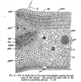
Leaf structure of Parsonsia heterophylla.png 856 × 862; 655 KB
-
Mesophyte leaf (34321748034).jpg 3,264 × 1,840; 1.05 MB
-
Mesophyte leaf (34321749754).jpg 2,609 × 1,470; 842 KB
-
Mesophyte leaf (34357209113).jpg 3,264 × 1,840; 1.12 MB
-
Mesophyte leaf (34810761446).jpg 3,264 × 1,840; 819 KB
-
Mesophyte x hydrophyte leaf (34381760130).jpg 3,264 × 1,840; 1.28 MB
-
Mesophytic Leaf Cross Section Microscope Image.jpg 1,280 × 960; 300 KB
-
NeriumLeafCross.png 4,000 × 3,000; 18.95 MB
-
Nymphaea leaf cross-section.jpg 1,424 × 1,512; 1.13 MB
-
Picture Natural History - No 316 - Section of Leaf.png 499 × 364; 371 KB
-
Pter1026 fern leaf L.jpg 1,200 × 736; 71 KB
-
Pter1026 fern leaf.jpg 1,200 × 736; 68 KB
-
Příčný řez kořenem Juncus articulatus.jpg 4,080 × 3,072; 2.34 MB
-
Rust fungi on Alnus glutinosa leaf surface.tif 1,023 × 669; 669 KB
-
Salt glands of the mangrove species Avicennia officinalis.png 1,248 × 762; 1.98 MB
-
Saxifraga oppositifolia - coupes des feuilles.jpg 1,842 × 2,272; 224 KB
-
Saxifraga stem.jpg 2,040 × 1,536; 1.19 MB
-
Sclerids embedded in upper epidermis of hydrophyte leaf (35268633765).jpg 3,264 × 1,840; 1.25 MB
-
Sclerids in lower epidermis of hydrophyte leaf (35228410636).jpg 3,264 × 1,840; 1.27 MB
-
Sclerids in the hydrophyte leaf (35268634785).jpg 3,264 × 1,840; 1.09 MB
-
Sclerids in upper epidermis of hydrophyte (35228405986).jpg 3,264 × 1,840; 1.43 MB
-
Sclerids support in the lower epidermis of a hydrophyte leaf (35268636095).jpg 3,264 × 1,840; 1.22 MB
-
Scleroid (35228407326).jpg 3,264 × 1,840; 1.06 MB
-
Scleroid in epidermis of hydrophyte leaf (34810755436).jpg 3,264 × 1,840; 1.07 MB
-
Scleroid in lower epidermis of hydrophyte leaf (34686901272).jpg 3,264 × 1,840; 741 KB
-
Scleroid support in the hydrophyte leaf (35228408416).jpg 3,264 × 1,840; 1.37 MB
-
SEM0041.TIF 660 × 500; 979 KB
-
The anatomy of some desert plants (1915) (18190954152).jpg 2,207 × 2,886; 825 KB
-
The anatomy of some desert plants (1915) (18195746131).jpg 2,637 × 3,314; 1.49 MB
-
The Oak (Marshall Ward) Fig 22.jpg 1,430 × 1,303; 137 KB
-
Tomato leaf x-section 2.jpg 1,024 × 1,049; 199 KB
-
Trachid and vessels within xylem bundle of mesophyte leaf (34605001012).jpg 3,264 × 1,840; 1.12 MB
-
Tradescantia, leaf, Etzold green 1.JPG 2,592 × 1,728; 2.88 MB
-
Tradescantia, leaf, Etzold green 2.JPG 1,792 × 2,688; 3.59 MB
-
Water Hyacinth Lamina cross section.jpg 1,600 × 1,200; 433 KB
-
Xerophyte leaf x mesophyte leaf (34381760970).jpg 3,264 × 1,840; 1.44 MB
-
Xerophytic Leaf Cross Section Stained Microscope Slide.jpg 1,280 × 960; 307 KB
-
Поперечный срез листа.png 2,592 × 1,944; 8.11 MB
-
Поперечный срез побега Casuarina equisetifolia.jpg 3,543 × 2,362; 4.96 MB
-
Поперечный срез столбчатого мезофила листа огурца (вместе с эпидермисом).jpg 5,501 × 5,761; 6.23 MB
-
Срез побега казуарины.jpg 3,543 × 2,362; 4.82 MB


