Category:Microtubules
Jump to navigation
Jump to search
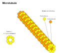
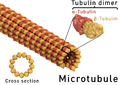
hollow tubes of internal diameter 12-15 nm and external diameter 24 nm found in a wide variety of eukaryotic cells | |||||
| Upload media | |||||
| Instance of |
| ||||
|---|---|---|---|---|---|
| Subclass of |
| ||||
| Part of |
| ||||
| Has part(s) |
| ||||
| |||||
Subcategories
This category has the following 10 subcategories, out of 10 total.
Media in category "Microtubules"
The following 50 files are in this category, out of 50 total.
-
De-Mikrotubulus.ogg 2.3 s; 22 KB
-
 201704 microtubule.svg 512 × 410; 573 KB
-
 ATP駆動型バイオマシーン(改4).jpg 1,026 × 993; 368 KB
-
 C21orf58 Subcellular Localization.png 1,366 × 1,190; 2.39 MB
-
 Comparison of bacterial and eukaryotic microtubules.jpg 390 × 824; 228 KB
-
 Crowded cytosol.png 2,308 × 1,988; 9.72 MB
-
 DESIGN (Figure3).jpg 1,280 × 720; 72 KB
-
 DESIGN(Figure1).jpg 1,280 × 720; 60 KB
-
 DESIGN(Figure2).jpg 1,280 × 720; 79 KB
-
 Dynamic instability.svg 562 × 509; 154 KB
-
 Dynamic microtubules are necessary for tail retraction in migrating cell.jpg 1,720 × 1,360; 229 KB
-
 Dynamiczna niestabilność mikrotubul007.PNG 957 × 342; 5 KB
-
 Embryo in flower.png 3,000 × 3,006; 2.97 MB
-
 Formation of Microtubule.png 3,152 × 1,475; 121 KB
-
 Fragment of Sus scrofa microtubule stabilized with taxol and peloruside.jpg 9,171 × 9,196; 8.24 MB
-
 HIF transzláció.jpg 876 × 508; 99 KB
-
 Journal.pbio.1001213.g004.png 980 × 824; 423 KB
-
 Mecanisme d'acció KM1.jpg 731 × 451; 52 KB
-
 Mecanisme d'acció Pyr1.jpg 462 × 577; 52 KB
-
 MicroT.png 1,780 × 1,063; 719 KB
-
 Microtubule id.svg 624 × 298; 40 KB
-
 Microtubule in Cell Migration.jpg 1,720 × 1,360; 230 KB
-
 Microtubule structure esp.png 2,200 × 1,535; 1.9 MB
-
 Microtubule structure Ukr.png 2,200 × 1,554; 1.74 MB
-
 Microtubule Structure-fr.svg 512 × 454; 184 KB
-
 Microtubule structure.png 2,200 × 1,554; 1.53 MB
-
 Microtubule Structure.svg 512 × 454; 176 KB
-
 Microtubule.png 793 × 771; 1.11 MB
-
 Microtubule.svg 624 × 298; 96 KB
-
 Microtubules and alkaloids pl.png 1,414 × 720; 85 KB
-
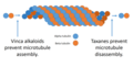 Microtubules and alkaloids.png 1,414 × 720; 117 KB
-
 Microtubules corr.svg 2,100 × 1,150; 316 KB
-
 Microtubules.png 432 × 329; 50 KB
-
 Microtubules.svg 2,100 × 1,150; 402 KB
-
 Microtubuli completo.jpeg 1,392 × 900; 357 KB
-
 Microtubulos.jpg 944 × 333; 93 KB
-
 Microtúbulo. Dinamica-fr.png 973 × 660; 197 KB
-
 Microtúbulo. Dinamica.png 1,005 × 681; 149 KB
-
 Mikrotub.svg 200 × 300; 865 KB
-
 Mikrotubula007 de.png 800 × 272; 3 KB
-
 Mikrotubula007 en.png 800 × 272; 3 KB
-
 Mikrotubula007.PNG 812 × 278; 3 KB
-
 Mikrotubulus kötőhelyek.jpg 364 × 434; 84 KB
-
 MT nomal.png 2,260 × 714; 267 KB
-
 Polimerizzazione Microtubuli.jpg 2,000 × 1,855; 443 KB
-
 ProteineTau ita.jpg 312 × 264; 20 KB
-
 ProteineTau.jpg 312 × 264; 23 KB
-
 Schematic of Cell State Splitter Organelle.jpg 324 × 215; 14 KB
-
 微小管の配向(英語).png 1,691 × 842; 1,011 KB
-
 微小管の配向.png 1,691 × 842; 875 KB

.jpg)


.jpg)
.jpg)
.jpg)


.png)
