Category:Mikael Häggström/Medical image annotations
Jump to navigation
Jump to search

Medical images annotated by Mikael Häggström[edit]
Media in category "Mikael Häggström/Medical image annotations"
The following 92 files are in this category, out of 92 total.
-
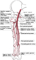
 Arteria et vena femoralis communis et subsartorialis.png 3,069 × 1,802; 1.23 MB
-
 Bohler's angle (raster).jpg 990 × 750; 139 KB
-
 Bohler's angle (vector).svg 198 × 150; 223 KB
-
 Caput-sourcil angle.jpg 1,143 × 704; 106 KB
-
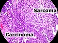 Carcinosarcoma of the endometrium - annotated.jpg 2,048 × 1,536; 1.17 MB
-
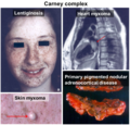 Carney complex with main associated diseases.png 865 × 829; 1.07 MB
-
 Common femoral and subsartorial artery and vein.jpg 2,931 × 1,806; 498 KB
-
 Coronary vessels, with annotated arteries.svg 894 × 573; 190 KB
-
 CT of the ostiomeatal complex, coronal plane, no annotations.png 1,081 × 579; 204 KB
-
 CT of the ostiomeatal complex, coronal plane, with annotations.png 2,165 × 1,380; 680 KB
-
 CT of the ostiomeatal complex, coronal plane, with annotations.svg 1,081 × 579; 319 KB
-
 Cytology of acute promyelocytic leukemia, annotated.png 1,393 × 943; 1.08 MB
-
 Decoy cell cytology.png 939 × 541; 436 KB
-
 Decoy cell on HE stain.png 372 × 356; 122 KB
-
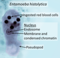 Entamoeba histolytica trophozoite.png 695 × 680; 427 KB
-
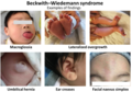 Examples of findings in Beckwith–Wiedemann syndrome.png 1,399 × 979; 1.22 MB
-
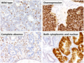
 Expression of p53 in urothelial neoplasms.png 856 × 924; 725 KB
-
 Fictional depiction of erythrocyte antigen neutralizers.png 957 × 509; 870 KB
-
 Gray84 clean.png 300 × 252; 42 KB
-
 HER2 FISH with a yellow signal.png 585 × 419; 181 KB
-
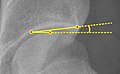
 Hip-knee-ankle angle.png 235 × 1,145; 106 KB
-
 Histology of gastric chief cell.png 92 × 98; 15 KB
-
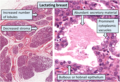 Histology of lactating breast, annotated.png 1,223 × 833; 1.92 MB
-
 Histology of lactating breast.jpg 1,227 × 837; 471 KB
-
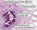 Histopathology of calcifying aponeurotic fibroma.png 783 × 643; 1,013 KB
-
 Histopathology of cholesterolosis, with annotated foam cell.jpg 704 × 526; 142 KB
-
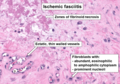 Histopathology of ischemic fasciitis.png 835 × 585; 854 KB
-
 Histopathology of microinvasive ductal carcinoma in situ.png 466 × 387; 454 KB
-
 Histopathology of myxoid liposarcoma.png 528 × 451; 445 KB
-
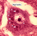 Histopathology of Negri bodies in rabies encephalitis.png 873 × 841; 1.24 MB
-
 Histopathology of proliferative fasciitis or myositis.png 465 × 372; 239 KB
-
 Histopathology of serrations in a traditional serrated adenoma.jpg 335 × 521; 66 KB
-

-
 HKA and HKS angles.svg 66 × 322; 235 KB
-
 Human anatomy planes, labeled.jpg 4,280 × 3,696; 914 KB
-
 HV and IM angles of hallux valgus.jpg 801 × 2,097; 313 KB
-
 HV and IM angles of hallux valgus.svg 801 × 2,097; 1.41 MB
-

-

-
 Image quality checking of pediatric pelvis.jpg 1,040 × 569; 201 KB
-
 Insall-Salvati ratio of patella baja.jpg 878 × 938; 120 KB
-
 Insall-Salvati ratio of patella baja.svg 878 × 938; 169 KB
-
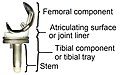 Knee prosthesis components.jpg 985 × 616; 125 KB
-
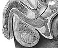 Lateral cross-section of male reproductive system.jpg 1,456 × 1,187; 517 KB
-
 Lateral cross-section of prostate.jpg 1,475 × 1,249; 506 KB
-
 Lateral cross-section of prostate.svg 1,475 × 1,250; 4.18 MB
-
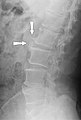 Lateral X-ray of lumbar spine spondylosis.jpg 1,431 × 2,116; 476 KB
-
 Leg length measurement on X-ray.jpg 1,557 × 4,247; 1,006 KB
-
 Leg length measurement on X-ray.svg 778 × 2,124; 1.78 MB
-
 Lobules and ducts of the breast.jpg 1,542 × 1,835; 736 KB
-
 Main antinuclear antibody patterns on immunofluorescence.png 814 × 596; 417 KB
-
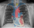
 Mediastinal structures on chest X-ray, annotated.jpg 2,573 × 2,086; 1.16 MB
-
 Mediastinal structures on chest X-ray.svg 2,573 × 2,086; 4.28 MB
-

-
 Micrograph of early actinic keratosis with parakeratosis.jpg 417 × 889; 99 KB
-
 Micrograph of homogenization of collagen.jpg 278 × 179; 22 KB
-

-
 Microscopic lymph node screening.jpg 1,341 × 957; 568 KB
-
 Non-chicken lymphoma.png 1,251 × 720; 1.11 MB
-
 Nuclear grooves.jpg 196 × 163; 14 KB
-
 Parietal cell and chief cells.png 413 × 441; 337 KB
-
 Parietal cell.png 91 × 95; 15 KB
-
 Patterns of p53 expression.png 945 × 711; 1.36 MB
-
 Progression of pancreatic intraepithelial neoplasia.png 974 × 344; 481 KB
-
 Proximal fractures of 5th metatarsal-es.svg 854 × 886; 3.2 MB
-
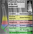 Proximal fractures of 5th metatarsal.jpg 1,709 × 1,772; 666 KB
-
 Proximal fractures of 5th metatarsal.svg 854 × 886; 3.18 MB
-
 Scaphoid fracture classification.jpg 545 × 614; 70 KB
-
 Schwannoma with Antoni A and Antoni B areas.jpg 604 × 465; 140 KB
-
 Sgarlato's angle of metatarsus adductus.jpg 652 × 1,127; 172 KB
-
 Sgarlato's angle of metatarsus adductus.svg 652 × 1,127; 199 KB
-
 Spondylolisthesis measurement on X-ray.png 390 × 411; 52 KB
-
 Spondylolisthesis measurement on X-ray.svg 195 × 206; 257 KB
-
 Subacromial space on outlet view X-ray.jpg 217 × 225; 22 KB
-
 The Internet denialist teacher.png 1,561 × 1,409; 2 MB
-
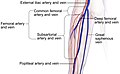
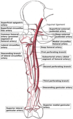 Thigh arteries.png 732 × 1,195; 435 KB
-
 Thigh arteries.svg 348 × 569; 78 KB
-
 Ultrasonography of keyhole sign of lower urinary tract obstruction.jpg 1,063 × 913; 133 KB
-
 Urine crystals comparison.png 1,129 × 977; 1.19 MB
-
 Vacuolar interface dermatitis, annotated.jpg 205 × 194; 22 KB
-
 Vertebra cervicalis.png 1,165 × 538; 192 KB
-
 Vertebra cervicalis.svg 546 × 252; 75 KB
-
 Viral infections and involved species-es.png 1,024 × 979; 379 KB
-
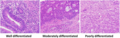 Well-, moderately and poorly differentiated colorectal adenocarcinoma.png 1,772 × 560; 2.05 MB
-
 X-ray of dynamic scapholunate instability - annotated.jpg 847 × 690; 190 KB
-
 X-ray of flexion angle of proximal interphalangeal joint of second toe.jpg 1,317 × 1,229; 300 KB
-

-
 X-ray of knee prosthesis with overhang.jpg 947 × 1,317; 236 KB
-
 X-ray of scoliosis, annotated.png 221 × 669; 144 KB
-
 X-ray of supraspinatus calcified tendinopathy, labeled.jpg 569 × 657; 85 KB
-
 X-ray of vertebral lines.jpg 1,285 × 1,465; 458 KB
-
 X-ray of vertebral lines.svg 890 × 1,014; 158 KB

.jpg)
.svg)


