Category:Neurons
Jump to navigation
Jump to search
electrically excitable cell that communicates via synapses   | |||||
| Upload media | |||||
| Instance of | |||||
|---|---|---|---|---|---|
| Subclass of |
| ||||
| Part of | |||||
| Has part(s) | |||||
| Significant event |
| ||||
| Different from | |||||
| |||||
Subcategories
This category has the following 34 subcategories, out of 34 total.
*
?
A
C
- C-fibres (1 F)
G
- GABAergic neurons (6 F)
K
- Koniocellular cell (2 F)
N
- Neural stem cells (147 F)
- Neurites (25 F)
- Neuritis (12 F)
- Neuron migration (22 F)
- Neuronal morphology (100 F)
- Number of neurons (5 F)
P
R
- Rattus neurons (10 F)
S
- Single neuron function (15 F)
W
Media in category "Neurons"
The following 200 files are in this category, out of 275 total.
(previous page) (next page)-
 10 caffeine knowing-neurons.jpg 838 × 1,024; 80 KB
-
 11 hemisphere knowing-neurons.jpg 838 × 1,024; 61 KB
-
 11-dimension.jpg 720 × 987; 224 KB
-
 12035 2012 8320 Fig7 HTML.webp 609 × 449; 80 KB
-
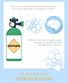 13 oxygen knowing-neurons3.jpg 838 × 1,024; 85 KB
-
 14 conduction knowing-neurons1.jpg 838 × 1,024; 71 KB
-
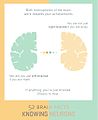
 15 intelligence knowing-neurons.jpg 838 × 1,024; 61 KB
-
 17 earth knowing-neurons.jpg 838 × 1,024; 86 KB
-
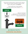 18 thalamus knowing-neurons1.jpg 838 × 1,024; 101 KB
-
 202101 Interneuron.svg 512 × 512; 899 KB
-
 202101 Motor neuron.svg 512 × 512; 1.01 MB
-
 202101 Three types of neurons.svg 512 × 512; 1.75 MB
-
 3D Medical Animation Still of Neuron.png 905 × 540; 520 KB
-
3D-реконструкция среза коры головного мозга крысы in situ.webm 6.7 s, 1,502 × 928; 719 KB
-
 4 sinapse orgasmo.jpg 2,048 × 2,048; 1.78 MB
-
 4 spill 2 knowing-neurons2.jpg 838 × 1,024; 84 KB
-
 4-week-old neurons (7184654817).jpg 2,048 × 2,048; 1.57 MB
-
 6 pain knowing-neurons.jpg 838 × 1,024; 74 KB
-
 7 bbb knowing-neurons.jpg 838 × 1,024; 117 KB
-
 8 serotonin 2 knowing-neurons1.jpg 838 × 1,024; 68 KB
-
 9 trifle 2 knowing-neurons.jpg 838 × 1,024; 94 KB
-

-

-
 A synapse between neurons.png 472 × 500; 173 KB
-
-
 A17 амакринові клітиини сітківки.png 1,222 × 885; 418 KB
-

-
 Action P7.jpg 1,120 × 550; 172 KB
-
 Alzheimers Disease.jpg 3,300 × 2,550; 1.82 MB
-
 Alzheimers neurons-tangles-bundles.PNG 637 × 337; 386 KB
-
-
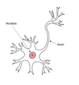
 Anatomy of neuron.png 1,849 × 1,037; 347 KB
-
 AP decay.png 408 × 485; 96 KB
-
 Artificialneuronhjdevias.jpeg 1,536 × 1,187; 192 KB
-
-
 Axo-axonic synapse.svg 512 × 601; 23 KB
-
 Axon of a neuron.jpg 960 × 540; 114 KB
-
 Axon Propagation.svg 512 × 1,050; 1.61 MB
-
 Axon Segmento Initial.png 1,099 × 1,530; 1.07 MB
-
 AxonTurning.jpg 1,806 × 899; 349 KB
-
 BAC firing.gif 718 × 480; 275 KB
-
 Back propagating action potential.gif 718 × 480; 109 KB
-
 Bastonetes e células bipolares no claro e no escuro eletrofisiologia.png 1,360 × 768; 91 KB
-
 Bastonetes e células bipolares no claro e no escuro simples.png 1,188 × 1,083; 98 KB
-
 Beta-Amyloid Plaques and Tau in the Brain (38686503251).png 1,920 × 1,080; 3.15 MB
-
 Biological neuron.gif 432 × 246; 4 KB
-
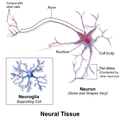
 Blausen 0672 NeuralTissue esp.png 2,000 × 2,000; 876 KB
-
 Blausen 0672 NeuralTissue ku.png 763 × 655; 190 KB
-
 Blausen 0672 NeuralTissue zh.png 2,000 × 2,000; 1.06 MB
-
 Blausen 0672 NeuralTissue.png 2,000 × 2,000; 1.04 MB
-
BN1 Neurology.ogv 5 min 28 s, 1,280 × 720; 53.13 MB
-
 Brain Chip (46771920474).jpg 674 × 526; 104 KB
-
 BruceBlausen Neuron RU.png 2,646 × 1,612; 1.05 MB
-
 Burst101.gif 478 × 170; 7 KB
-
 Bursting Examples.gif 409 × 468; 11 KB
-
 Bursting-recording.png 661 × 197; 27 KB
-
 Ca spike.gif 718 × 480; 234 KB
-
 CA1 pyramidal cells with synapses.png 455 × 894; 277 KB
-
 Cajal Discussion.JPG 3,264 × 2,448; 2.08 MB
-
 Cajal-Retzius cell drawing by Cajal 1891.gif 440 × 93; 12 KB
-
 Canais de sodio e potassio despolarizacao hiperpolarizacao.png 1,418 × 770; 140 KB
-
 Candelabro 03 CUATRIS.jpg 377 × 445; 128 KB
-
 Cell types in human frontal agranular insular cortex.png 2,002 × 2,341; 895 KB
-
 Celula del lobulo cerebral electrico del torpedo. Wellcome L0040800.jpg 2,664 × 2,870; 2.06 MB
-
 Celula gigante de la porcion inferior del asta de ammon Wellcome L0040799.jpg 2,632 × 3,528; 2.34 MB
-
 Cerebrosterol.jpg 517 × 427; 60 KB
-

-
 Channels.png 310 × 203; 24 KB
-
 Chemical synapse schema cropped az.jpg 481 × 599; 205 KB
-
 Chk-UCH1-GFAP-20X-1.jpg 1,600 × 1,200; 3.07 MB
-
Chopin Etude op 10 n 3.oga 3 min 36 s; 1.66 MB
-
-
-
-
-
-
-
-
-
-
-
-
-
-
-
-
-
 Components of neuron.jpg 2,046 × 1,412; 461 KB
-
 Control compuerta A activado.png 793 × 661; 72 KB
-
 Control compuerta sin A.png 789 × 665; 72 KB
-
 Convolutional Neural Network NeuralNetworkFeatureLayers.gif 960 × 720; 127 KB
-
 Convolutional Neural Network NeuralNetworkFilter.gif 960 × 720; 96 KB
-
 Convolutional Neural Network with Color Image Filter.gif 960 × 720; 129 KB
-
 Convolutional Neural Network.gif 960 × 720; 50 KB
-
 CPCA-GFAP,MCA-5B10,Tau,neurons.jpg 1,000 × 1,000; 921 KB
-
 Célula Nervosa Impulsos.PNG 624 × 600; 69 KB
-
 DANs Art.jpg 945 × 945; 558 KB
-
De-Nervenzelle.ogg 2.2 s; 21 KB
-
De-Neuron.ogg 1.8 s; 16 KB
-
-
 Demystifying Depression-Neurons.png 540 × 257; 13 KB
-
 Dendritic ap spike.gif 474 × 318; 9 KB
-
 Description fonctionnelle du fonctionnement d'un neurone.png 593 × 280; 8 KB
-
 Differentiation of Stem Cells Into Neurons.jpg 1,884 × 835; 845 KB
-
Digital Neuron Museum - interactive digital catalog visualizing structural and functional data for mouse retinal cell types.webm 1 min 46 s, 2,304 × 1,440; 90.91 MB
-
 Diseases of the nervous system (1910) (14586295439).jpg 1,616 × 1,808; 185 KB
-
 Dissecting Neurons.jpg 4,752 × 3,168; 4.24 MB
-
 DPI Silicon Neuron.jpg 416 × 532; 104 KB
-
 DrPaulineNeveu 01 NeuroneBipolaire.svg 827 × 397; 44 KB
-
 DrPaulineNeveu 01 NeuroneMultipolaire.svg 772 × 703; 45 KB
-
 DrPaulineNeveu 01 NeuronePseudoUnipolaire.svg 716 × 260; 9 KB
-
 DrPaulineNeveu 02 Recepteurs senso nue encaps cell.png 960 × 812; 143 KB
-
 DrPaulineNeveu 06 UM.svg 854 × 421; 27 KB
-
 DrPaulineNeveuNeuroneEpineDendritique.svg 716 × 324; 30 KB
-
 DrPaulineNeveuNeuroneMultipolaireGaineMyéline.svg 677 × 314; 25 KB
-
 Dying neurons (4541410256).jpg 1,737 × 1,737; 2.03 MB
-
-

-
 Evolutionäre spezifizierung von zellen zu nervenz.jpg 651 × 222; 29 KB
-
 Example of a neuron.png 1,402 × 774; 626 KB
-
 Fasern.jpg 889 × 781; 154 KB
-
 Fig5-Critic-Actor-Synapses.png 1,176 × 816; 85 KB
-
 Figura1.Marcha.jpg 1,449 × 1,581; 365 KB
-
 Figura2.Marcha.jpg 1,481 × 1,657; 705 KB
-
 Figura3.Marcha.jpg 646 × 856; 80 KB
-
 Figura4.Marcha.jpg 1,225 × 856; 196 KB
-
 Figura5.Marcha.jpg 1,005 × 721; 159 KB
-
 Figure 35 01 02.png 1,043 × 700; 339 KB
-
 Figure 35 01 03.jpg 1,117 × 515; 218 KB
-
 Figure 35 01 05.jpg 544 × 431; 161 KB
-
 Figure 35 01 06.jpg 1,117 × 544; 430 KB
-
 Figure 35 01 07.jpg 1,117 × 522; 270 KB
-
 Figure 35 04 01f.png 1,044 × 1,386; 194 KB
-
 Forced burst.gif 451 × 121; 5 KB
-
 From clusteron to sigmoidal neuron.png 746 × 1,063; 61 KB
-
 Frontal insula.png 274 × 361; 50 KB
-
 Ganglion cell.jpg 1,440 × 1,440; 220 KB
-
 GFP Neurons.png 2,317 × 1,992; 6.04 MB
-
 GFP TH staining of Mouse Primary Culture Neuron.jpg 606 × 606; 159 KB
-
 Glial cells ku.png 907 × 551; 369 KB
-
 GnRH Neuron.png 343 × 438; 352 KB
-
 Histology of thalamic neuron.jpg 2,048 × 1,532; 431 KB
-
 Human Cell Groups distributed by Cell Count and by Aggregate Cell Mass.jpg 3,162 × 2,096; 1.08 MB
-
 Human Cell Groups; Cell Count, Cell Mass, and Aggregate Cell Mass (Biomass).png 1,258 × 847; 149 KB
-
 Human neuron.jpg 2,992 × 4,000; 2.95 MB
-
 Hypothesized Propagation of Activity in Human Neocortex.jpeg 611 × 538; 26 KB
-
 Impulsfortleitung an der Nervenzelle.png 987 × 949; 35 KB
-
 Interneuron releasing neuromodulators.jpg 1,295 × 767; 125 KB
-
 Intrinsic bursting2.gif 240 × 297; 5 KB
-
 Isopotential contours for a spherical soma with 7 cylindrical dendrites.png 1,448 × 1,387; 342 KB
-
 Jul0709 C4 MCH.jpg 5,748 × 2,622; 551 KB
-
 Known age-related electrophysiological and morphological changes in neurons.jpg 950 × 1,454; 579 KB
-

-
 Layer V Pyramidal cell from mouse visual cortex.png 1,075 × 838; 244 KB
-
 Le neurone, une cellule spécialisée.jpg 590 × 560; 63 KB
-
 Magnetogenetic manipulation of neuron.png 966 × 468; 15 KB
-
 Major metabolic fluxes in neuron-astrocyte coupling for resting conditions.png 1,024 × 710; 330 KB
-
 MCA-6H112 NFL 4K citra HuCbl 20X 02-wiki.jpg 1,920 × 1,440; 1.66 MB
-

-
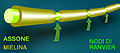
 Meyeline.png 1,309 × 431; 532 KB
-
 Meyers b6 s0894 b1.png 333 × 433; 20 KB
-
 MINFLUX image of a hippocampal neuron.jpg 827 × 565; 71 KB
-
 Model of inhibitory neurogenesis.png 711 × 520; 152 KB
-
 Modified Axo-axonic synapse.svg 482 × 482; 42 KB
-

-
 Mouse brain cells.jpg 715 × 715; 148 KB
-
 Mouse Dorsal Root Ganglion.tif 998 × 958; 2.77 MB
-
 Myelin sheath damage in multiple sclerosis.svg 985 × 767; 91 KB
-
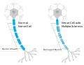
 Myelinated unmylinated neurons.PNG 1,300 × 1,300; 55 KB
-
 Nerve cell from anterior horn of spinal cord grey matter Wellcome L0002018.jpg 1,092 × 1,690; 345 KB
-
 Nerve cell with "Endfusse" on it. Wellcome L0002907.jpg 1,166 × 1,648; 286 KB
-
 Nerve cell.png 639 × 686; 62 KB
-
 Nerve cells of various kinds. Wellcome L0002007.jpg 1,261 × 1,407; 210 KB
-
 Nerve copy.jpg 614 × 272; 72 KB
-
 Nervengewebe.jpg 944 × 1,352; 1.98 MB
-
 Nervenzellelp.png 284 × 491; 36 KB
-
 Nervios raquideos.jpg 214 × 144; 8 KB
-
 Neural cell.jpg 1,609 × 988; 639 KB
-
Neural Nets 3200-25.ogv 5 min 37 s, 1,280 × 720; 44.97 MB
-
-
-
-
 Neuro-stub.png 60 × 44; 7 KB
-
 Neurococi.png 816 × 967; 67 KB
-
Neurology Engrammes.ogv 5 min 11 s, 1,280 × 720; 87.73 MB
-
 Neuron (action potential) illustration.webp 591 × 498; 67 KB
-
 Neuron (Turkish).jpg 13,068 × 7,280; 9.25 MB
-
 Neuron 011910.JPG 4,080 × 3,072; 4.04 MB
-
 Neuron icon.png 47 × 83; 5 KB
-
 Neuron in the microchannel of hydrogel implant.png 5,240 × 1,034; 9.97 MB
-
 Neuron sections.gif 606 × 160; 2 KB
-
 Neuron Structure.png 600 × 323; 67 KB
-
 Neuron togopic.png 400 × 436; 63 KB
-
 Neuron with ALL channels.jpg 1,498 × 654; 137 KB
-
 Neuron-matrix.png 800 × 600; 330 KB
-
 Neuron-section.jpg 1,660 × 1,694; 245 KB
-
 Neuron-SEM-2.png 800 × 600; 114 KB
-
 Neuron-SEM.png 800 × 600; 219 KB
-
 Neuron-tech.png 800 × 600; 257 KB
-
 Neuron-toon.png 800 × 600; 102 KB


.jpg)


_(14763266295).jpg)


.png)


.jpg)


_(14586295439).jpg)


.jpg)


.png)


.png)


_illustration.webp)
.jpg)


