Category:Nucleosides
Jump to navigation
Jump to search
glycosylamine that can be thought of as nucleotide without a phosphate group  | |||||
| Upload media | |||||
| Instance of |
| ||||
|---|---|---|---|---|---|
| Subclass of |
| ||||
| Part of |
| ||||
| |||||
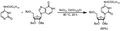
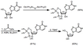
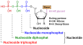
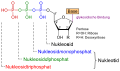
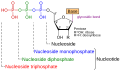
English: Nucleosides are molecules consisting of a nucleobase linked to a five-carbon sugar. A nucleoside with one or more phosphate group is called a nucleotide. Nucleosides are glycosylamines made by attaching a nucleobase to a ribose ring. Examples of these include cytidine, uridine, adenosine, guanosine, thymidine and inosine.
Subcategories
This category has the following 39 subcategories, out of 39 total.
A
- Azacitidine (7 F)
- Azaribine (3 F)
- Azvudine (2 F)
C
- Cytidine (14 F)
- Cytidine derivatives (16 F)
D
- Decitabine (6 F)
- Deoxyadenosine (7 F)
- Deoxycytidine (8 F)
- Deoxyguanosine (6 F)
- Deoxythymidine (11 F)
- Deoxyuridine (6 F)
- Dihydrouridine (3 F)
E
- EdU (4 F)
F
- Fazarabine (3 F)
- Filociclovir (2 F)
G
I
- Immucillin H (6 F)
M
- Maribavir (2 F)
- N1-Methylpseudouridine (1 F)
- Molnupiravir (14 F)
N
- Nicotinamide riboside (4 F)
P
- Pentostatin (6 F)
- Pseudouridine (10 F)
Q
- Queuosine (2 F)
R
- Ribavirin (7 F)
- Ribothymidine (5 F)
S
T
- Taribavirin (3 F)
U
Z
Media in category "Nucleosides"
The following 100 files are in this category, out of 100 total.
-
 00 Nucleoside.png 323 × 171; 11 KB
-
 18F-flt (structural formula).png 380 × 600; 16 KB
-
 7 nukleozid kinázok.png 816 × 437; 12 KB
-
 7DMA.svg 168 × 228; 45 KB
-
 Aminoallyl Uridine labled.jpg 508 × 388; 12 KB
-
 Archaeosine Archaeosin.svg 620 × 528; 12 KB
-
 BCX4430 structure.png 610 × 292; 20 KB
-
 Building nucleic acids SVG.svg 512 × 388; 74 KB
-
 Carbocyclic Nuc & Bases.svg 23,470 × 7,466; 190 KB
-
 Cf1743.png 1,879 × 524; 18 KB
-
 CGS21680.png 848 × 581; 12 KB
-
 Clitidine 01.svg 615 × 420; 13 KB
-
 Clitocine.svg 512 × 515; 19 KB
-
 CMX521 structure.png 1,187 × 839; 39 KB
-
 Convergent and Linear Synthesis of Carbocyclic Nucleosides.svg 19,846 × 5,383; 85 KB
-
 D5SICS-skeletal.png 801 × 1,109; 45 KB
-
 D5SICS.png 490 × 257; 57 KB
-
 Deoxyadenosyl radical.svg 140 × 128; 17 KB
-
 Deoxyinosine.png 483 × 565; 21 KB
-
 Deoxyribonucleoside generic.png 780 × 642; 29 KB
-
 Deoxyxanthosine.png 1,200 × 1,218; 41 KB
-
 Deuremidevir.svg 760 × 585; 40 KB
-
 DNaM-skeletal.png 890 × 969; 45 KB
-
 DNaM.svg 625 × 375; 12 KB
-
 Edge of Interaction on Nucleoside.png 1,112 × 1,094; 113 KB
-
 EICAR structure.png 1,381 × 853; 45 KB
-
 EICAR.svg 645 × 400; 14 KB
-
 Floxuridin alovudin zidovudin.svg 332 × 420; 33 KB
-
 Formation of 8oxodG.png 612 × 180; 15 KB
-
 Funcionament dels de derivats de nucleòsids.png 724 × 467; 36 KB
-
 Funcionament dels derivats de nucleòsids.png 649 × 449; 35 KB
-
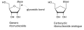 Generic ribonucleoside with gycosidic bond and carbocyclic ribonucleoside analogue.svg 12,881 × 4,551; 87 KB
-
 GS-6620 structure.png 929 × 759; 44 KB
-
 IDX-184 structure.png 1,491 × 826; 51 KB
-
 KP-1461 metabolism and activation.svg 1,668 × 1,198; 103 KB
-
 KP-1461.svg 695 × 492; 9 KB
-
 Loxoribine.svg 188 × 230; 20 KB
-
 Lumicitabine structure.png 690 × 851; 37 KB
-
 Lysidine A.png 768 × 1,296; 42 KB
-
 Mec. de acció DN.png 527 × 400; 30 KB
-
 MECA.svg 700 × 280; 26 KB
-
-
 MK-608 structure.png 442 × 348; 16 KB
-
 MK-608.svg 675 × 270; 17 KB
-
 N-MCT structure.png 1,295 × 809; 42 KB
-
 Namodenoson.svg 940 × 365; 35 KB
-
 NITD008.svg 675 × 270; 18 KB
-
 Nucl Amidite Arbuzov.png 437 × 125; 4 KB
-
 NucleoAlt1.png 509 × 168; 22 KB
-
 NucleoAlt2.png 749 × 164; 28 KB
-
 NucleoEx.png 623 × 175; 25 KB
-
 NucleoGen1.png 541 × 161; 19 KB
-
 NucleoGen2.png 542 × 147; 17 KB
-
 NucleoGen3.png 580 × 167; 22 KB
-
 NucleoMech.png 761 × 492; 58 KB
-
 NucleoScope1.png 571 × 191; 25 KB
-
 NucleoScope2.png 559 × 158; 20 KB
-
 NucleoScope3.png 611 × 170; 27 KB
-
 NucleoScope4.png 641 × 168; 24 KB
-
 NucleoScope5.png 614 × 341; 42 KB
-
 Nucleoside biochemistry.svg 512 × 512; 41 KB
-
 Nucleoside nucleotide general format.png 2,183 × 1,184; 73 KB
-
 Nucleoside Phosphitylation 1.png 1,413 × 359; 14 KB
-
 Nucleoside Phosphitylation 2.png 1,413 × 359; 13 KB
-
 Nucleoside Phosphoramidite Arbuzov Reaction.png 1,221 × 416; 11 KB
-
 Nucleoside Phosphoramidite Oxidation.png 2,155 × 446; 20 KB
-
 Nucleoside Phosphoramidite Reactivity.png 2,880 × 445; 25 KB
-
 Nucleoside phosphoramidites.png 2,115 × 999; 34 KB
-
 Nucleoside Salvage Pathway.png 234 × 694; 25 KB
-
 Nucleoside.png 1,242 × 990; 20 KB
-
 Nucleotide nucleoside general (hun).png 1,138 × 747; 55 KB
-
 Nucleotide nucleoside general bg.svg 820 × 484; 50 KB
-
 Nucleotide nucleoside general it.svg 820 × 484; 51 KB
-
 Nucleotide nucleoside general vi.svg 877 × 484; 157 KB
-
 Nucleotide nucleoside general-de.svg 820 × 484; 105 KB
-
 Nucleotide nucleoside general-tr.svg 820 × 484; 35 KB
-
 Nucleotide nucleoside general.svg 820 × 484; 46 KB
-
 Nucleotides 1 FR.svg 820 × 290; 180 KB
-
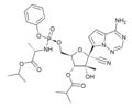
 Nukleozyd z grupami ochronnymi.jpg 2,191 × 2,078; 365 KB
-
 Obeldesivir.svg 740 × 395; 28 KB
-
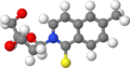
 Pentostatin ball-and-stick.png 3,000 × 1,692; 572 KB
-
 Phosphoramidite.png 1,024 × 856; 22 KB
-
 Prepared Carbocyclic Nucleoside Analogs by Using Vince Lactam.JPG 826 × 355; 38 KB
-

-

-
 PSI-6130.png 1,621 × 809; 20 KB
-
 Pyrazofurin.svg 645 × 400; 14 KB
-
 Ribonucleoside generic.png 780 × 642; 30 KB
-
 Sangivamycin structure.png 1,189 × 748; 37 KB
-
 Sangivamycin.svg 675 × 400; 17 KB
-
 Sapacitabine.png 1,778 × 591; 21 KB
-
 Structure of CCPA.png 1,841 × 802; 21 KB
-
 T-1106.svg 605 × 420; 14 KB
-
 Tectitethya crypta in Medicine.png 631 × 510; 381 KB
-
 Tiazofurin.svg 620 × 618; 7 KB
-
 Triciribine.svg 1,155 × 910; 19 KB
-
 Valopicitabine structure.png 1,187 × 799; 40 KB
-
 Wikifig.tif 3,904 × 3,158; 265 KB
-
 Wybutosine.svg 620 × 537; 16 KB
-
 Wyosine - Wyosin.svg 620 × 416; 15 KB

.png)


.png)


