Category:Orthopantomograms
Jump to navigation
Jump to search
Deutsch: Ein Orthopantomogramm ist eine diagnostische Röntgenaufnahme der Ober- und Unterkiefer des Menschen. Es werden alle Zähne, die angrenzenden Kieferbereiche, beide Kiefergelenke und ebenso die rechte und die linke Kieferhöhle abgebildet.
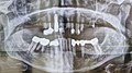
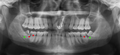
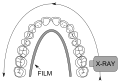
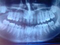
English: A panoramic radiograph is a panoramic scanning dental X-ray of the upper and lower jaw. It shows a two-dimensional view of a half-circle from ear to ear.
dental radiogram depicting the maxilla, or both maxilla and mandible, on a single film | |||||
| Upload media | |||||
| Subclass of |
| ||||
|---|---|---|---|---|---|
| |||||
Media in category "Orthopantomograms"
The following 116 files are in this category, out of 116 total.
-
 X- ray with ear ring.jpg 3,584 × 2,213; 3.35 MB
-
 1471-2350-8-52-2-l.jpg 571 × 518; 35 KB
-
 Orthopantomogram of an abscessed tooth.jpg 2,960 × 1,440; 251 KB
-
 2022-07-11 Gebiss eines Neunzigjährigen, Röntgenaufnahme.jpg 3,704 × 2,083; 1.74 MB
-
 ApikaleOstitis4.JPG 720 × 576; 37 KB
-
 Asymptomatic disease free impacted wisdom teeth.jpg 3,076 × 1,542; 1.65 MB
-
 Basic panoramic radiograph.jpg 2,843 × 1,384; 321 KB
-
 Cleidocranial dysplasia teeth.jpg 1,145 × 537; 74 KB
-
 Computed tomography slice showing impacted teeth.jpg 1,099 × 783; 194 KB
-
 Cyst - wisdom tooth.jpg 1,537 × 861; 123 KB
-
 Deep Inside MariPeru.jpg 2,176 × 1,010; 139 KB
-
 Dent001.JPG 1,006 × 581; 61 KB
-

-
 Dental Panorama X-ray.jpg 2,048 × 1,088; 297 KB
-
 Dental X-rays.JPG 2,857 × 1,376; 1.45 MB
-
 DenticiónMixta.jpg 400 × 281; 11 KB
-
 DenticiónMixta1.jpg 400 × 256; 8 KB
-
 Dentition2.JPG 720 × 576; 35 KB
-
 Dentition3.JPG 720 × 576; 39 KB
-
 Dentition4.JPG 720 × 576; 42 KB
-
 Dentition5.JPG 720 × 576; 42 KB
-
 Doctor Catalán Bajuelo.jpg 300 × 200; 26 KB
-
 DVT-scan-MagoCut-bild1-singlesample.jpg 500 × 500; 40 KB
-
 DVT-scan-MagoCut-bild2-oversamplefocus.jpg 500 × 500; 30 KB
-
 Eckzahn retiniert und verlagert Zahn 13 OPG 20100106 001.JPG 4,272 × 2,848; 2.89 MB
-
 Eckzahn retiniert und verlagert Zahn 13 OPG 20100106 002.JPG 4,272 × 2,848; 2.8 MB
-
 Eckzahn retiniert und verlagert Zahn 13 OPG 20100106 003.JPG 4,272 × 2,848; 3.17 MB
-
 Florid osseous dysplasia.png 956 × 403; 316 KB
-
 Generalized perio -touched up.jpg 871 × 325; 29 KB
-
 Hyperdontie Röntgen.jpg 703 × 304; 46 KB
-
 Impacted wisdom teeth caries.png 2,626 × 1,212; 1.91 MB
-
 Impacted wisdom teeth.jpg 2,617 × 1,244; 140 KB
-
 Jaw xray.jpg 954 × 885; 398 KB
-
 Melketenner og jeksler IMG 9580.jpg 3,524 × 2,158; 4.3 MB
-
 Melketenner og jeksler IMG 9581.jpg 3,519 × 2,214; 4.51 MB
-
 Milk-tooth and impacted inverted 2nd premolar.jpg 3,084 × 1,560; 1.23 MB
-
 Numata panorama xray teeth.svg 4,557 × 3,134; 65 KB
-
 OPG Aufnahme.jpg 1,557 × 786; 348 KB
-
 OPG IMG 6681.jpg 4,794 × 2,291; 4.31 MB
-
 OPG IMG 6683.jpg 5,616 × 3,744; 8.79 MB
-
 OPG IMG 6684.jpg 5,616 × 3,744; 11.39 MB
-
 OPG IMG 6685.jpg 5,616 × 3,744; 10.85 MB
-
 OPG IMG 7570.JPG 4,272 × 2,848; 2.71 MB
-
 OPG of condylar atrophy.png 1,242 × 725; 621 KB
-
 OPG w56J -.jpg 1,612 × 856; 203 KB
-
 Opg-2015-02-04.jpg 2,840 × 1,531; 430 KB
-
 OPG-Bild.jpg 543 × 251; 28 KB
-
 Opg-mask.jpg 2,000 × 1,616; 280 KB
-
 Opg.png 1,440 × 900; 757 KB
-
 Orthopanthomogram.jpg 1,224 × 697; 213 KB
-
 Orthopantogram3.jpg 2,985 × 1,244; 348 KB
-
 Orthopantomogram Bloch-Sulzberger syndrome.jpg 553 × 281; 23 KB
-
 Orthopantomogram of a patient with missing lower incisors.jpg 1,952 × 1,024; 542 KB
-
 Orthopantomogram of missing eight side wisdom teeth in upper and lower arches.jpg 1,952 × 1,024; 615 KB
-
 Orthopantomogram of moderate crowding.jpg 1,935 × 1,024; 571 KB
-
 Orthopantomogram of severe dental crowding.jpg 1,935 × 1,024; 566 KB
-
 Orthopantomogram showing impacted upper canine.jpg 1,952 × 1,024; 532 KB
-
 Orthopantomogram showing mesio angular impaction of third molars.jpg 1,952 × 1,024; 655 KB
-
 Orthopantomogram showing rotated central incisors.jpg 1,935 × 1,024; 554 KB
-
 Orthopantomogram showing supra erupted third molar.jpg 1,935 × 1,024; 608 KB
-
 Orthopantomogram.jpg 1,048 × 510; 530 KB
-
 Orthopantomograms.jpg 2,000 × 1,050; 218 KB
-
 Orthopantomogram̠ showing supernumerary teeth.jpg 1,952 × 1,024; 705 KB
-
 ORTO 1.jpg 1,709 × 1,066; 127 KB
-
 ORTO 2 RecortadaPS.jpg 746 × 416; 69 KB
-
 ORTO 2.jpg 1,814 × 982; 143 KB
-
 Ortopantomografia (OPT).jpg 1,753 × 891; 276 KB
-
 Ortopantomografia.jpg 700 × 437; 31 KB
-
 Ortopantomografía.jpg 1,406 × 738; 272 KB
-
 Ortopantomogram popis.jpg 500 × 259; 48 KB
-
 Ortopantomogram.jpg 500 × 259; 15 KB
-
 ORTORecortadaPS Medias lunas azules.jpg 746 × 416; 72 KB
-
 PAN TEETH.jpg 2,816 × 1,540; 1.23 MB
-
 Panoramaröntgen.tiff 6,247 × 3,376; 27.36 MB
-
 Panoramic braces.jpg 1,736 × 837; 194 KB
-
 Panoramic radiograph - Orthopantomogram.jpg 3,292 × 1,536; 2.22 MB
-
 Panoramic radiograph of historic dental implants.jpg 1,350 × 904; 98 KB
-
 Panoramicfilm.JPG 2,816 × 1,540; 1.2 MB
-
 PanoramicRXhoriz.jpg 604 × 302; 32 KB
-
 Panoramicxray.JPG 2,268 × 1,422; 458 KB
-
 Panorex radiograph from a patient with dentinogenesis imperfecta.jpg 444 × 263; 12 KB
-
 Pantomog dntindysplasia.jpg 1,200 × 597; 96 KB
-
 Radiografía bucal.jpg 2,592 × 1,542; 533 KB
-
 RadiografíaDientes.jpg 500 × 245; 17 KB
-
 Roentgenaufnahme gebiss 20040920.jpg 2,948 × 1,552; 342 KB
-
 Roentgenschuerze.png 744 × 1,196; 519 KB
-
 Rontgen foto gebit.jpg 1,668 × 724; 46 KB
-
 RotatingPan.gif 231 × 100; 96 KB
-
 Simple mandible fracture.jpg 2,692 × 1,341; 1.53 MB
-
 Subperiostal Implants 20100105 000.JPG 4,240 × 2,528; 4.27 MB
-
 Subperiostal Implants 20100105 001.JPG 4,272 × 2,848; 2.71 MB
-
 Subperiostal Implants 20100105 002.JPG 2,848 × 4,272; 2.74 MB
-
 Subperiostal Implants 20100105 003.JPG 2,848 × 4,272; 3.15 MB
-
 Subperiostal Implants 20100105 005.JPG 4,258 × 2,632; 4.35 MB
-
 Subperiostal Implants 20100105 006.JPG 4,272 × 2,848; 2.91 MB
-
 Subperiostal Implants 20100105 007.JPG 4,272 × 2,848; 3.01 MB
-
 Subperiostal Implants 20100105 008.JPG 4,272 × 2,848; 2.75 MB
-
 Teeth front X ray.jpg 640 × 480; 35 KB
-

-
 Wechselgebiss OPG Ausschnitt 20100106 001.JPG 4,272 × 2,848; 3.15 MB
-
 Wechselgebiss OPG Ausschnitt 20100106 002.JPG 4,272 × 2,848; 2.71 MB
-
 Wechselgebiss OPG Ausschnitt 20100106 003.JPG 4,272 × 2,848; 2.79 MB
-
 Wechselgebiss OPG Ausschnitt 20100106 004.JPG 4,272 × 2,848; 3.01 MB
-
 Wechselgebiss1.jpg 720 × 576; 44 KB
-
 Wechselgebiss2.jpg 720 × 576; 41 KB
-
 Weisheitszahn10.JPG 1,600 × 857; 648 KB
-
 Weisheitszahn5.JPG 720 × 576; 37 KB
-
 Weisheitszahn6.JPG 720 × 576; 34 KB
-
 Weisheitszahn7.JPG 720 × 576; 31 KB
-
 Weisheitszahn8.JPG 720 × 576; 32 KB
-
 Weisheitszahn9.JPG 720 × 576; 32 KB
-
 X-ray of all 32 human teeth.jpg 2,606 × 1,244; 256 KB
-
 X-ray of teeth.jpg 2,867 × 1,536; 1.69 MB
-
 X-ray64.JPG 1,522 × 826; 151 KB
-
 X-ray64A.JPG 381 × 207; 13 KB
-
 Xray of four impacted teeth.jpg 2,592 × 1,944; 319 KB


.jpg)


.jpg)


.jpg)