Category:Primitive node
Jump to navigation
Jump to search
Media in category "Primitive node"
The following 49 files are in this category, out of 49 total.
-
ADMP restricts the size of the organizer domain Spemann's organizer.jpg 818 × 665; 397 KB
-
ALK1 is required to restrict the size of the organizer domain Spemann's organizer.jpg 1,374 × 1,026; 1.27 MB
-
Aves Dorsal view of a twenty-five-hour chick embryo with seven primitive segments.jpg 1,993 × 1,744; 2.21 MB
-
Aves FGF signalling in mesoderm migration.jpg 2,152 × 612; 371 KB
-
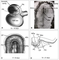
Characterization of the SOX2T-positive territory of the epiblast in chicken embryo.jpg 2,113 × 1,581; 1.21 MB
-
Cilia are present and functional in the node of 4HH-8HH talpid3 chickens.jpg 1,063 × 1,402; 582 KB
-
Crown-cells-sense-nodal-flow-through-Pkd2-a-Pkd2-expression-throughout-the-node-pit.jpg 1,251 × 1,259; 523 KB
-
De Novo Formation of Left–Right Asymmetry by Posterior Tilt of Nodal Cilia.ogv 13 s, 320 × 240; 2.05 MB
-
Diagrams and images of human embryos at the gastrula stage.png 3,128 × 3,193; 804 KB
-
Dose-dependent reshaping of primitive streak.jpg 968 × 1,236; 743 KB
-
Embryo disc human.jpg 3,326 × 2,096; 449 KB
-
Flow Generated by the Mechanical Model cilia primitive node.ogv 9.9 s, 320 × 240; 77 KB
-
Flow Generation Mechanism of cilia primitve node.png 2,020 × 1,451; 238 KB
-
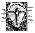
Formation of notochord by primitive streak.jpg 1,091 × 1,071; 167 KB
-
Hans Spemann.jpg 849 × 1,371; 500 KB
-
Leftward Flow Generated near the Surface.ogv 17 s, 360 × 240; 515 KB
-
Localization of PGCs in mouse, rabbit and chick by the end of gastrulation.png 1,204 × 931; 587 KB
-
Migration of epiblast cells in the mammalian embryo.png 866 × 530; 281 KB
-
Models to explain the function of nodal flow in L–R asymmetry.jpg 1,279 × 1,155; 600 KB
-
Node Cilia Are Posteriorly Tilted and Positioned primitive node.png 2,020 × 3,157; 4.01 MB
-
Notopterus notopterus (10.3897-zse.93.13341) Figure 10.jpg 1,961 × 2,062; 2.04 MB
-
Number of TSOX2 double-positive cells during early chicken stages embryo.jpg 2,007 × 2,502; 828 KB
-
Outline of key signaling pathways.png 4,148 × 1,682; 1.3 MB
-
Pathway of visceral left–right determination in the vertebrate.png 3,614 × 1,774; 572 KB
-
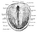
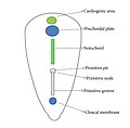
Primitiv Node.jpg 720 × 504; 42 KB
-
Quantification of the number of epiblast cells electroporated chicken embryo.jpg 730 × 1,687; 273 KB
-
Sus domesticus embryos The primitive streak.jpg 1,123 × 912; 408 KB
-
The Biological bulletin (20187079648).jpg 1,854 × 2,206; 1.25 MB
-
The left-right asymmetry pathway mouse embryo.jpg 1,691 × 893; 432 KB
-
Trajectory of Node Cilia Movement.png 2,800 × 1,728; 2.48 MB
-
Types of cilia found at the left-right-organizer of vertebrates.jpg 1,027 × 1,309; 719 KB


