Category:Receptor proteins
Jump to navigation
Jump to search
Wikimedia category | |||||
| Upload media | |||||
| Instance of | |||||
|---|---|---|---|---|---|
| |||||
Subcategories
This category has the following 36 subcategories, out of 36 total.
7
A
- Amino acid receptors (14 F)
- Aryl hydrocarbon receptor (10 F)
C
- CCR7 receptors (17 F)
E
- Epidermal growth factor receptor (92 F)
G
- GABA receptor (6 F)
I
- IgG receptors (10 F)
L
M
- Membrane receptor signaling (99 F)
N
- Neuropeptide Y receptors (7 F)
S
T
- Thyrotropin receptors (9 F)
- Transferrin receptors (5 F)
V
- Vitronectin receptors (2 F)
Media in category "Receptor proteins"
The following 65 files are in this category, out of 65 total.
-
 1hg4.png 282 × 266; 75 KB
-
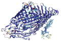
 1ten fibronectin type III.png 720 × 1,146; 249 KB
-
 201601 Receptor.png 400 × 268; 69 KB
-
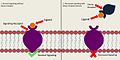
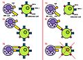
 Activation vs silencing of PAR.png 917 × 747; 54 KB
-
 Agonists v2.png 2,349 × 1,146; 45 KB
-
 ASGP-R.svg 768 × 860; 9 KB
-
 Asgpr (2).jpg 640 × 761; 112 KB
-
 Blodtrycksreglering.svg 512 × 581; 305 KB
-
 BMPR1A.png 486 × 440; 58 KB
-
 C5a-receptor.png 960 × 720; 77 KB
-
 COXII-receptor.png 1,326 × 1,261; 309 KB
-
 CTLA4 Crystal Structure.jpg 500 × 700; 76 KB
-
 CTLA4 Crystal Structure.rsh.png 739 × 750; 244 KB
-
 Decoy Receptor Figure-raster.png 1,152 × 651; 228 KB
-
 DecoyReceptorFigure.jpg 720 × 360; 72 KB
-
 EGF Receptor.jpg 720 × 540; 30 KB
-
 EphA5.png 360 × 430; 84 KB
-
 Erb fig.jpeg 1,038 × 550; 114 KB
-
 Fas receptor.png 3,200 × 2,400; 1.84 MB
-
 Fc receptor schematic big.png 600 × 600; 10 KB
-
 FcAr.png 600 × 600; 14 KB
-
 FhuA.png 540 × 370; 198 KB
-
 HLA-G Receptors.jpg 1,589 × 1,124; 285 KB
-
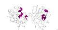
 Human RAGEvC1.png 1,700 × 990; 258 KB
-
 IFNAR Ligand Binding.png 827 × 423; 34 KB
-
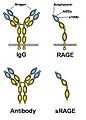 IgG-RAGE.jpg 541 × 756; 146 KB
-
 Imaoaction.JPG 926 × 515; 99 KB
-
 KAR fosforilation.jpg 742 × 732; 44 KB
-
 KAR imagen.jpg 1,276 × 923; 138 KB
-
 LDLR 1N7D.png 1,701 × 1,776; 710 KB
-
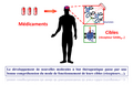 Les récepteurs membranaires comme cibles thérapeutiques.png 3,184 × 2,050; 163 KB
-

-
 Membrane Receptors.svg 512 × 288; 101 KB
-
 Neuropilin.png 1,436 × 759; 246 KB
-
 Nozizeptor.jpg 720 × 540; 49 KB
-
 PBB Protein ESR1 image.png 960 × 720; 369 KB
-
 PBB Protein INSR image.jpg 500 × 500; 35 KB
-
 PBB Protein NR1H4 image.jpg 500 × 500; 36 KB
-
 PBB Protein NR1I3 image.jpg 500 × 500; 56 KB
-
 PBB Protein NR3C2 image.jpg 500 × 500; 27 KB
-
 PBB Protein NTRK1 image.jpg 500 × 500; 44 KB
-
 PBB Protein RARA image.jpg 500 × 500; 51 KB
-
 PBB Protein RARB image.jpg 500 × 500; 33 KB
-
 PBB Protein RXRA image.jpg 500 × 500; 34 KB
-
 PBB Protein THRA image.jpg 500 × 500; 41 KB
-
 PBB Protein THRB image.jpg 500 × 500; 29 KB
-
 Pdgfrb.jpg 789 × 575; 30 KB
-
 PPAR-diagram-es.png 3,448 × 2,032; 400 KB
-
 PPAR-diagram.png 3,448 × 2,032; 2.72 MB
-
 Receptor Sigma-1.png 776 × 511; 140 KB
-
 Receptor02.jpg 334 × 286; 59 KB
-
 Receptors.jpg 546 × 220; 43 KB
-
 Rimonabant Pharmacophore.png 1,387 × 1,688; 54 KB
-
 Récepteurs tableau.jpg 1,664 × 1,023; 127 KB
-
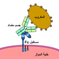
 Schéma-du-récepteur-Fc.png 600 × 558; 74 KB
-
 Signaltransduktion Geruchsrezeptor.png 833 × 374; 36 KB
-
 Starke alpha-Autorezeptor1972.jpg 2,540 × 1,160; 151 KB
-
 Steroid receptor.png 800 × 558; 125 KB
-
 Structure des récepteurs NOD1 et NOD2 et leurs ligands.jpg 580 × 362; 21 KB
-
 TGF beta pathway - step 1.svg 216 × 480; 5 KB
-
 TGFBeta step1.png 216 × 480; 15 KB
-
 TLR4.png 600 × 523; 271 KB
-
 Transferrin cycle.png 1,354 × 1,080; 573 KB
-
 TrkB-schema-eng.png 1,044 × 1,404; 76 KB
-
 Vreceptor.jpg 300 × 400; 25 KB


.jpg)


