Category:Scanning electron microscopic images of Protista
Jump to navigation
Jump to search
Subcategories
This category has the following 2 subcategories, out of 2 total.
Media in category "Scanning electron microscopic images of Protista"
The following 48 files are in this category, out of 48 total.
-
-
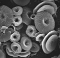
 9Calcidiscus leptoporus, diploid, SEM, showing coccoliths.tif 755 × 765; 721 KB
-
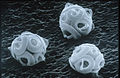
 Baculogypsina sphaerulata.jpg 512 × 512; 139 KB
-
 Calcarina sp.jpg 512 × 512; 157 KB
-
 CSIRO ScienceImage 4847 SEM dinoflagellate.jpg 1,858 × 1,209; 1.52 MB
-
 CSIRO ScienceImage 7202 SEM Coccolithophorid (1).jpg 832 × 832; 510 KB
-
 CSIRO ScienceImage 7202 SEM Coccolithophorid.jpg 1,861 × 1,202; 1.59 MB
-
 CSIRO ScienceImage 7596 dinoflagellate.jpg 1,861 × 1,202; 1.71 MB
-
 CSIRO ScienceImage 7608 Pyrodinium.jpg 1,827 × 1,220; 2.14 MB
-
 CSIRO ScienceImage 7609 SEM dinoflagellate.jpg 1,831 × 1,192; 2.07 MB
-
 CSIRO ScienceImage 8289 SEM diatom.jpg 1,861 × 1,217; 1.66 MB
-
 Cyclococcolithus-leptoporus hg.jpg 1,906 × 1,906; 461 KB
-
 Cyclococcolithus-leptoporus-back hg.jpg 2,072 × 2,072; 476 KB
-
 Detail, Cyclococcolithus-leptoporus hg (cropped).jpg 813 × 1,020; 130 KB
-
 Detail, Diatom algae Amphora sp (cropped).jpg 377 × 466; 180 KB
-
 Detail, Euglypha sp (cropped).jpg 173 × 217; 36 KB
-
 Detail, Neogloboquadrina pachyderma hg (cropped).png 215 × 269; 58 KB
-
 Diatom algae (Bacillariophyta).jpg 1,280 × 960; 1.25 MB
-
 Diatom algae Amphora sp.jpg 1,280 × 960; 1.11 MB
-
 Diatom algae and pollen.jpg 1,280 × 960; 1.03 MB
-
 Diatoms-HCMR.jpg 712 × 484; 251 KB
-
 Diatoms.png 1,400 × 1,144; 951 KB
-
 Emiliania huxleyi coccolithophore (PLoS).png 650 × 650; 301 KB
-
 Euglypha sp.jpg 640 × 480; 102 KB
-
 Gephyrocapsa oceanica brighter.jpg 1,600 × 1,600; 1.67 MB
-
 Gephyrocapsa oceanica color (lightened).jpg 1,600 × 1,600; 3.2 MB
-
 Gephyrocapsa oceanica color.jpg 1,600 × 1,600; 1.74 MB
-
 Gephyrocapsa oceanica.jpg 1,600 × 1,600; 933 KB
-
 Giardia lamblia in a late stage of cell division.jpg 648 × 768; 349 KB
-
 Isotricha intestinalis.jpg 2,000 × 1,489; 944 KB
-
 N pachyderma hg.png 653 × 451; 267 KB
-
 Nitzschia-kerguelensis hg.jpg 2,706 × 2,236; 1.5 MB
-
 Parasite180015-fig2 Sicuophora multigranularis (Armophorea, Clevelandellida).png 4,252 × 3,195; 8.28 MB
-
 Podomonas kaiyoae C.jpg 756 × 493; 156 KB
-
 SEM Euglypha sp.jpg 960 × 720; 181 KB
-
 Stephanopogon sp.jpg 640 × 480; 155 KB
-
 Synura asmundiae.jpg 5,120 × 3,840; 5.39 MB
-
 Synura petersenii.png 408 × 759; 261 KB
-
 Thoracosphaera-cocco hg.jpg 2,878 × 2,251; 1.25 MB
-
 Thoracosphaera-heimi-open hg.jpg 2,859 × 2,211; 1.29 MB
-
 Thoracosphaera-heimi2 hg.jpg 2,542 × 2,032; 496 KB
-
 Thoracosphaera-open hg.jpg 2,568 × 2,072; 611 KB
-
 Thoracosphaera-sp-diag hg.jpg 2,841 × 2,259; 1.28 MB
-
 Thoracosphaera2 hg.jpg 2,854 × 2,278; 1.03 MB
-
 Thorascosphaera-sp hg.jpg 2,587 × 2,073; 560 KB
-
 Thorascosphaere-bbb hg.jpg 2,536 × 2,031; 725 KB
-
 Динофитовая микроводоросль, выделенная из осадков Амурского залива в 2020 году.jpg 1,680 × 1,315; 457 KB
-
 Динофлагелляты Scrippsiella 2020 Amur bay.jpg 1,360 × 1,636; 610 KB


.jpg)


.jpg)
.jpg)
.jpg)
.png)
.jpg)


.png)


.jpg)


.png)


