Category:Sensory system
Jump to navigation
Jump to search
part of the nervous system responsible for processing sensory information | |||||
| Upload media | |||||
| Instance of |
| ||||
|---|---|---|---|---|---|
| Subclass of |
| ||||
| Part of | |||||
| Has part(s) | |||||
| |||||
Subcategories
This category has the following 41 subcategories, out of 41 total.
- SVG sensory system (16 F)
*
- Interoception (10 F)
A
D
E
- Electroreception (41 F)
F
- Sensory feedback (12 F)
G
H
M
- Mechanoreceptors (18 F)
N
- Neural adaptation (2 F)
P
- Prepulse inhibition (4 F)
R
- Receptive field (17 F)
S
- Sense organs (23 F)
- Spatial behavior (14 F)
- Startle reactions (14 F)
- Stimming (25 F)
V
Media in category "Sensory system"
The following 62 files are in this category, out of 62 total.
-
 1543,Vesalius'Fabrica,VisualSystem,V1.jpg 286 × 309; 25 KB
-
 1543,Visalius'OpticChiasma.jpg 287 × 308; 24 KB
-
 Abeille, pétiole.jpg 2,687 × 2,257; 267 KB
-
 Ampullae of Lorenzini.png 1,000 × 378; 12 KB
-
 Ampullae of Lorenzini.svg 960 × 720; 49 KB
-

-
 Ascending pain pathways.tif 1,563 × 1,812; 551 KB
-
 Blindspot.gif 365 × 28; 365 bytes
-
 Blindspot.jpg 300 × 50; 844 bytes
-

-
 Brodmann areas 17 18 19.png 256 × 192; 40 KB
-
 Challenge Modell.jpg 336 × 349; 64 KB
-
 ClawedFrogLarvaeUnidentified.JPG 2,272 × 1,704; 1.38 MB
-
 Continuous Interleaved Sampling.jpg 637 × 295; 32 KB
-
 Diseases of the nervous system (1910) (14770796114).jpg 1,748 × 2,088; 480 KB
-
 DISPOSITIVO DE AYUDA Y ORIENTACION VISUAL.pdf 1,275 × 2,100; 116 KB
-
 DISPOSITIVO DE ORIENTACION VISUAL.jpg 6,805 × 10,000; 6.5 MB
-

-
 Enose01.png 994 × 765; 24 KB
-
 Esquema activació indirecta.png 792 × 178; 8 KB
-
 EuropeanGardenSpiderAraneaDiadematus.JPG 2,272 × 1,704; 1.46 MB
-
 EyeFixationsReading.gif 597 × 130; 5 KB
-
 FAD reaction.png 1,163 × 544; 81 KB
-
 Fotomotor.jpg 747 × 582; 414 KB
-
 Four Types of Learning Styles.jpg 2,048 × 1,536; 125 KB
-
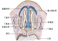 Gray51 zh.png 500 × 353; 124 KB
-
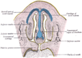 Gray51.png 500 × 353; 40 KB
-
 Illu cerebrum lobes.jpg 371 × 274; 34 KB
-
 Le traitement des informations sensorielles.jpg 1,905 × 1,107; 106 KB
-
 Le traitement des informations sensorielles.png 2,031 × 1,198; 126 KB
-
 Limiar e intensidade do sinal sensorial.png 1,528 × 543; 49 KB
-
 Lorenzini pores on snout of tiger shark.jpg 1,592 × 816; 108 KB
-
 Lorenzini Sensory Epithelium.png 734 × 790; 22 KB
-
 Lukt high sve.jpg 2,280 × 2,340; 1.52 MB
-
 Magnetite maghemite.png 1,049 × 415; 47 KB
-
 Mandibular nerve 1.jpg 960 × 720; 92 KB
-
 MuscleSpindle Model.png 800 × 278; 20 KB
-
 Neon colour spreading illusion.gif 434 × 444; 6 KB
-
 New Brunswick DGJ 4545 - Self Portrait (4281687677).jpg 4,120 × 2,580; 6.13 MB
-
 Nociceptive pain 2.jpg 504 × 724; 234 KB
-
 Nociceptive pain.jpg 1,920 × 1,080; 895 KB
-
 Noun Brain Nithinan 2452319.svg 512 × 512; 3 KB
-

-
 Prepulse Inhibition schematically rus.png 1,024 × 1,134; 156 KB
-
 Prepulse Inhibition schematically.png 1,024 × 768; 126 KB
-
 PushPull System.gif 300 × 228; 9 KB
-
 Receptive field sCH.png 403 × 737; 52 KB
-
 Receptive field-ar.png 403 × 737; 57 KB
-
 Receptive field.png 403 × 737; 67 KB
-
 Sensory cilium.jpg 649 × 296; 40 KB
-
 SensoryProcessing-ar.png 839 × 182; 83 KB
-
 SensoryProcessing.png 690 × 149; 16 KB
-
 Shark Orientation Sensing 1.png 1,242 × 776; 77 KB
-
 Shark Orientation Sensing 2.png 866 × 1,210; 53 KB
-
 SingleNeuron output.jpg 1,201 × 901; 63 KB
-
 Structure of sensory system (4 models) E.PNG 1,011 × 980; 142 KB
-
 Structure of sensory system (4 models) J.PNG 1,000 × 980; 138 KB
-
 Structure of sensory system (4 models) N.PNG 920 × 980; 115 KB
-
 Structure of sensory system (4 models)-ar.png 1,011 × 980; 163 KB
-
 Transducao.png 1,434 × 404; 21 KB
-
 Whiter than white illusion.jpg 453 × 223; 9 KB
-
 Дкг+.jpg 815 × 304; 32 KB


_(14770796114).jpg)


.jpg)


_(14778453101).jpg)


_E.PNG)
_J.PNG)
_N.PNG)
-ar.png)
