Category:Surgical suture
Jump to navigation
Jump to search
technique to close a surgical or traumatic wound | |||||
| Upload media | |||||
| Subclass of | |||||
|---|---|---|---|---|---|
| Part of | |||||
| |||||
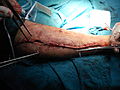
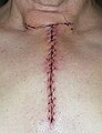
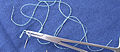
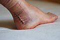
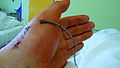
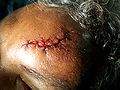
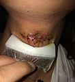
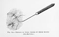
Sutures are the stitches doctors, and especially surgeons, use to hold skin, internal organs, blood vessels and all other tissues of the human body together, after they have been severed by injury or surgery.
Subcategories
This category has the following 8 subcategories, out of 8 total.
- SVG suture (32 F)
B
- Bone anchors (3 F)
D
- De hechtsteek (7 F)
H
K
- Surgeon's knot (1 P, 238 F)
O
S
W
- Sutured wounds (6 F)
Pages in category "Surgical suture"
This category contains only the following page.
Media in category "Surgical suture"
The following 167 files are in this category, out of 167 total.
-
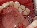
 2016-12-03 10-00-47 nodule (cropped).jpg 3,292 × 1,569; 1.4 MB
-
 2016-12-03 10-00-47 nodule.jpg 3,648 × 2,736; 2.2 MB
-
 2017 Kermanshah earthquake by Farzad Menati - Sarpol-e Zahab (52).jpg 800 × 532; 149 KB
-
 A Surgeon Applying a Suture.jpg 2,978 × 3,398; 1.38 MB
-
 After orthopedic surgery.JPG 4,000 × 3,000; 4.75 MB
-
 AfterThymectomy.JPG 353 × 459; 78 KB
-
 Ago montato.jpg 1,510 × 659; 390 KB
-
 Aller - Beim flicken.jpg 1,192 × 1,525; 881 KB
-
 Allers - Verbandstechniken.jpg 3,200 × 3,948; 3.2 MB
-
 An American text-book of the diseases of children (1895) (14782099782).jpg 1,824 × 2,160; 910 KB
-
 Annual and analytical cyclopaedia of practical medicine (1898) (14792915923).jpg 2,228 × 3,192; 1.52 MB
-

-
 Bear 1 third treatment sewing 1 (28050286159).jpg 3,264 × 2,448; 903 KB
-
 Bear 1 transport day last treatment 1 (39119473624).jpg 2,000 × 1,333; 1.87 MB
-
 Bear 1 transport day last treatment 2 (39828827541).jpg 2,000 × 1,333; 1.85 MB
-
 Bibliotheca anatomica, medica, chirurgica, etc Fleuron T113958-34.png 1,319 × 1,256; 75 KB
-
Buried knot suture technique.webm 1 min 52 s, 854 × 480; 10.92 MB
-
 Central Line Sutures.jpg 2,448 × 3,264; 1.21 MB
-
 Chirurgenknoten 01.svg 1,200 × 1,200; 4 KB
-
 Chirurgenknoten 02.svg 1,200 × 700; 4 KB
-
 Chirurgenknoten 03.svg 1,200 × 1,200; 4 KB
-
 Chirurgische Naht,Heilungsverlauf.jpg 1,008 × 336; 220 KB
-
 Chirurgische Naht2.jpg 800 × 533; 144 KB
-
 Clinical gyncology, medical and surgical (1895) (14761924916).jpg 1,418 × 870; 143 KB
-
 Closeup of repair.JPG 3,456 × 2,592; 3.21 MB
-
 Coated VICRYL.45cm.(SPATULA) 01.JPG 1,600 × 1,200; 961 KB
-
 Coated VICRYL.45cm.(SPATULA) 02.JPG 1,600 × 1,200; 942 KB
-
 Continous-Horizontal mattress suture.svg 560 × 440; 27 KB
-
Corner stitch suture technique.webm 1 min 45 s, 854 × 480; 10 MB
-
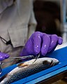
 Dave Smith fish-suturing collage (8595557989).jpg 931 × 864; 793 KB
-
 Diseases and disorders of the horse (Page 171) BHL23614341.jpg 2,309 × 3,869; 485 KB
-
 Diseases and disorders of the horse (Page 172) BHL23614342.jpg 2,309 × 3,869; 872 KB
-
-
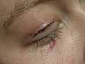 Eyelid's suture.jpg 2,816 × 2,112; 3.63 MB
-
 Gynaecology for students and practitioners (1916) (14779573394).jpg 1,608 × 1,904; 478 KB
-
 Hallus Valgus Bunion Cicatrice 8 jours.jpg 3,552 × 2,580; 1.44 MB
-
 Hallux valgus or bunion 03.jpg 4,416 × 2,560; 1.83 MB
-
 Hand Injury.jpg 1,374 × 1,158; 95 KB
-
 Hand with drain after surgery (1).jpg 3,712 × 2,088; 1.8 MB
-
 Hand with drain after surgery (2).jpg 3,712 × 2,088; 1.99 MB
-
 Head injury.jpg 3,472 × 2,604; 1.42 MB
-
 Hechtingen.JPG 3,072 × 2,304; 1.24 MB
-
 Histopathology of suture material.jpg 2,080 × 1,536; 970 KB
-
 Horizontal mattress suture.svg 560 × 440; 42 KB
-
 Implanon removed stitches.jpg 2,448 × 3,264; 897 KB
-
 Infected site.jpg 2,448 × 2,675; 1.54 MB
-
 Joe.JPG 1,280 × 960; 206 KB
-
 Joint Readiness Training Center 140117-F-XL333-869.jpg 4,256 × 2,832; 4.27 MB
-

-
 Lister, Excision wound stitched, dressed, Antiseptic Surgery Wellcome M0007804.jpg 4,114 × 2,792; 4.28 MB
-
 Lister, Method of tying vessels...Cheyne, Antiseptic Surgery Wellcome M0007798.jpg 4,143 × 2,613; 1.55 MB
-
 Lister, Stitched wound...Cheyne, Antiseptic Surgery. Wellcome M0007802.jpg 3,652 × 3,156; 6.07 MB
-
 Man with cheek wound; surgeon sews head wound, 14th C Wellcome L0037333.jpg 4,072 × 3,920; 4.05 MB
-
 NEEMO 7 McKinley suturing.jpg 3,040 × 2,008; 2.43 MB
-
 Operative gynecology (1898) (14781468411).jpg 1,350 × 1,482; 178 KB
-
 Operative gynecology - (1906) (14760598936).jpg 1,462 × 1,888; 760 KB
-
 Operative gynecology - (1906) (14781158524).jpg 1,318 × 1,574; 543 KB
-
 Operative gynecology - (1906) (14803371643).jpg 2,006 × 1,594; 730 KB
-
 Operative surgery (1899) (14593763687).jpg 1,038 × 1,688; 129 KB
-

-
 Operative surgery, for students and practitioners (1913) (14779371895).jpg 1,658 × 1,808; 793 KB
-
 Painless.jpg 2,592 × 1,944; 2.27 MB
-
 Pathology and treatment of diseases of women (1912) (14758654596).jpg 1,906 × 1,840; 1.05 MB
-
 Peque20210729 ohs01.jpg 1,216 × 1,280; 274 KB
-
 Peque20210729 ohs02.jpg 1,280 × 909; 214 KB
-
 Peque20210729 ohs03.jpg 1,095 × 1,280; 159 KB
-
 Peque20210729 ohs04.jpg 1,137 × 1,280; 269 KB
-
 Peque20210729 ohs05.jpg 996 × 1,280; 183 KB
-
 Peque20210729 ohs06.jpg 521 × 1,280; 36 KB
-
 Peque20210729 ohs07.jpg 1,194 × 1,280; 230 KB
-
 Peque20210729 ohs08.jpg 1,280 × 1,197; 90 KB
-
 Peque20210729 ohs09.jpg 1,280 × 1,008; 82 KB
-
 Peque20210729 ohs11.jpg 1,280 × 1,224; 309 KB
-
 Plaie doigt 01.jpg 1,344 × 1,008; 156 KB
-
 Plaie doigt 02.jpg 1,344 × 1,008; 143 KB
-
 Plaie doigt 03.jpg 1,344 × 1,008; 106 KB
-
 Plaie doigt 04.jpg 1,344 × 1,008; 134 KB
-
 Plaie doigt 05.jpg 1,344 × 1,008; 102 KB
-
 Plaie doigt 06.jpg 1,344 × 1,008; 145 KB
-
 Plaie suturée surinfectée.jpg 393 × 390; 43 KB
-
 Pocket surgical instrument set, London, England, 1846-1901 Wellcome L0057738.jpg 2,832 × 4,256; 1.64 MB
-
 Portraiture for a cloven lip Wellcome L0016286.jpg 1,182 × 1,604; 461 KB
-

-
 Punto di Donati.jpg 474 × 155; 44 KB
-
 Punto di sutura.jpg 500 × 178; 43 KB
-
 Rutschknoten 01.svg 1,200 × 1,600; 6 KB
-
 Rutschknoten 02.svg 1,200 × 1,600; 6 KB
-
 Rutschknoten 03.svg 1,200 × 1,600; 6 KB
-
 Rutschknoten 04.svg 1,200 × 1,600; 7 KB
-
 Rutschknoten 05.svg 1,200 × 1,600; 6 KB
-
 Scar makeup stitches.jpg 1,440 × 2,560; 1.12 MB
-
 Scar on hand after 18 days.jpg 2,440 × 1,680; 1.53 MB
-
 Scar on hand after 22 days.jpg 3,023 × 1,852; 1.83 MB
-
 Scars 6 days post surgery.JPG 1,632 × 1,224; 353 KB
-
 Schnittwunde am Knie nach Entfernung des Nahtmaterials.JPG 2,816 × 2,112; 2.01 MB
-
 Sebvarró fonal és tű 1a.jpg 539 × 534; 65 KB
-
 Shouldice Operation Fig. 1.jpg 4,755 × 4,527; 3.95 MB
-
Simple interrupted stitch suture technique.webm 1 min 54 s, 854 × 480; 5.3 MB
-
Slip knot suture technique with instruments.webm 1 min 29 s, 854 × 480; 4.09 MB
-
Square knot suture technique with instruments.webm 1 min 1 s, 854 × 480; 2.97 MB
-
Square knot suture technique with one hand.webm 53 s, 854 × 480; 2.64 MB
-
Square knot suture technique with two hands.webm 48 s, 854 × 480; 1.9 MB
-
 Stanislaus Operationsnarbe Weichteilsarkom 01.jpg 4,000 × 3,000; 9.21 MB
-
 Stanislaus Operationsnarbe Weichteilsarkom 02.jpg 3,000 × 4,000; 8.91 MB
-
 Steri-Strip - 01.jpg 1,024 × 768; 545 KB
-
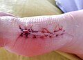
 Stitched finger.JPG 4,000 × 3,000; 2.28 MB
-
 Stitches 2.jpg 1,307 × 1,393; 212 KB
-
 Stitches post appendicitis surgery.jpg 2,816 × 2,112; 2.02 MB
-
 Surgical drains.jpg 4,248 × 5,664; 4.76 MB
-
 Surgical instrument set, Germany, 1939 Wellcome L0058116.jpg 4,068 × 2,832; 1.72 MB
-
 Surgical stiches - knee.JPG 2,816 × 2,112; 2.07 MB
-
 Surgical stitches.jpg 2,336 × 3,378; 3.48 MB
-
 Surgical suture knot001.svg 1,600 × 2,200; 42 KB
-
 Surgical suture.jpg 2,704 × 1,576; 688 KB
-
 Surgical thread supplier 1.jpg 1,051 × 1,576; 400 KB
-
 Sutura ad U orizzontale.jpg 761 × 663; 68 KB
-
 Sutura del pollice.jpg 1,280 × 960; 187 KB
-
 Sutura di Dufourmentel.jpg 500 × 163; 44 KB
-
 Sutura intradermica.jpg 761 × 663; 67 KB
-
 Suture - Landscaping Accident.jpg 600 × 800; 43 KB
-
Suture and suture needle holding technique.webm 54 s, 854 × 480; 2.66 MB
-
 Suture on knee.jpg 2,000 × 1,984; 1.35 MB
-
 Sutured wound.jpg 1,728 × 2,592; 1.13 MB
-
 SuturedFinger2.jpg 320 × 240; 34 KB
-
 SutureOut - Feature 1.jpg 5,184 × 3,456; 4.69 MB
-
 SutureOut - Sharps Container.jpg 3,456 × 5,184; 4.44 MB
-
 Sutures (15721902653).jpg 4,272 × 2,840; 2.67 MB
-
 Sutures 7.JPG 4,000 × 3,000; 2.57 MB
-
 Sutures a patients hand.jpg 1,400 × 1,000; 138 KB
-
 Sutures.jpg 2,592 × 1,221; 212 KB
-
 Sutures6.JPG 4,000 × 3,000; 2.21 MB
-
 SuturesFinger.jpg 480 × 360; 91 KB
-
 SuturesRemoved1.jpg 320 × 240; 39 KB
-
 The breast- its anomalies, its diseases, and their treatment (1917) (14570379069).jpg 1,440 × 1,336; 690 KB
-
 The breast- its anomalies, its diseases, and their treatment (1917) (14570452298).jpg 1,444 × 1,060; 573 KB
-
 The breast- its anomalies, its diseases, and their treatment (1917) (14570634677).jpg 1,444 × 1,124; 542 KB
-
 The breast- its anomalies, its diseases, and their treatment (1917) (14754693084).jpg 1,446 × 1,212; 639 KB
-
 The breast- its anomalies, its diseases, and their treatment (1917) (14754695134).jpg 1,440 × 1,032; 427 KB
-
 The breast- its anomalies, its diseases, and their treatment (1917) (14754725074).jpg 1,442 × 1,822; 790 KB
-
-
-
-
 The science and art of midwifery (1897) (14740590016).jpg 1,164 × 1,628; 141 KB
-
 Throwing Sutures Portrait (15721689183).jpg 2,690 × 3,363; 2.08 MB
-
 Tipo sutura.jpg 479 × 330; 58 KB
-
 Transactions of the Southern Surgical and Gynecological Association (1907) (14582881767).jpg 2,036 × 2,588; 1.07 MB
-
 Trépanation pour méningiome.JPG 400 × 300; 34 KB
-
 Tube of sterile catgut ligature, Edinburgh, Scotland, 1890-1 Wellcome L0058104.jpg 2,670 × 4,202; 1.31 MB
-

-

-

-

-

-

-

-

-

-
-

-
 Vasectomy.JPG 640 × 599; 234 KB
-
 VATS 03.jpg 1,181 × 886; 264 KB
-
 Ventriculoperitoneal shunt - surgical wound healing - belly - day 12.jpg 2,272 × 1,704; 1.53 MB
-

-
Vertical matress suture technique.webm 2 min 1 s, 854 × 480; 5.13 MB
-
 VICRYL.Violet braided absorbable suture.3-0.70cm.JPG 1,600 × 1,200; 972 KB
-
 Wundnaht bei offener Operationstechnik des Carpaltunnelsyndroms.jpg 2,048 × 1,536; 796 KB
-
 Техника хирургических швов.jpg 295 × 187; 8 KB
.jpg)

.jpg)


_(14782099782).jpg)
_(14792915923).jpg)
.jpg)


_(14761924916).jpg)

_01.JPG)
_02.JPG)

.jpg)
_BHL23614341.jpg)
_BHL23614342.jpg)


_(14779573394).jpg)


.jpg)
.jpg)


_(14781468411).jpg)
_(14760598936).jpg)
_(14781158524).jpg)
_(14803371643).jpg)
_(14593763687).jpg)
_(14578897799).jpg)
_(14779371895).jpg)

_(14758654596).jpg)


_(14784785482).jpg)


.jpg)


_(14570379069).jpg)
_(14570452298).jpg)
_(14570634677).jpg)
_(14754693084).jpg)
_(14754695134).jpg)
_(14754725074).jpg)
_(14595399199).jpg)
_(14763314104).jpg)
_(14785533793).jpg)
_(14740590016).jpg)
.jpg)


,10_v2.png)
,10.png)
,7.png)
,8.png)
,9.png)
.png)


_of_New_York,_a_Registered_Nurse_and_Clinical_administrator_for_the_Los_Angeles_County,_University_of_Southern_California.jpg)
_Medical_Center.jpg)


.jpg)
.jpg)
_(14582881767).jpg)