Category:T cells
Jump to navigation
Jump to search
type of lymphocyte  | |||||
| Upload media | |||||
| Instance of | |||||
|---|---|---|---|---|---|
| Subclass of | |||||
| Part of | |||||
| Named after | |||||
| |||||
Subcategories
This category has the following 19 subcategories, out of 19 total.
A
C
- CCR5 (4 F)
- CD3 (immunology) (3 F)
- CD8-positive T-lymphocytes (64 F)
H
- T helper cell (8 F)
- Human activated T-cells (8 F)
J
- Jurkat cells (69 F)
N
R
- T-cell receptor (7 F)
- Regulatory T-lymphocytes (10 F)
T
- T-lymphocyte subsets (7 F)
- Th1 cells (19 F)
- Th17 cells (8 F)
- Th2 cells (6 F)
V
Media in category "T cells"
The following 58 files are in this category, out of 58 total.
-
 202206 T lymphocyte.svg 512 × 512; 595 KB
-

-
 Advert for Guy's Tonic Wellcome L0040436.jpg 2,544 × 4,160; 3.09 MB
-
 Antigen presentation ar.svg 271 × 241; 204 KB
-
 APC Cross-Presentation.png 4,428 × 2,622; 1.26 MB
-
 Architektur eines chimären Antigenrezeptors (CAR).webp 2,055 × 939; 125 KB
-
 Blausen 0625 Lymphocyte T cell (crop).png 1,165 × 1,130; 1.71 MB
-
 Blausen 0625 Lymphocyte T cell.png 1,500 × 1,500; 1.41 MB
-
 Brady Microscopy.jpg 879 × 589; 356 KB
-
 CAR T-Cell Therapy.jpg 1,305 × 1,727; 626 KB
-
 CAR T-cell Therapy.svg 512 × 160; 74 KB
-
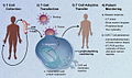 CAR-Engineered T-Cell Adoptive Transfer.jpg 1,280 × 758; 238 KB
-
 CDR and V,D, and J Genes.png 774 × 355; 29 KB
-
 Citoquinas.jpg 394 × 227; 53 KB
-
 Clonal Deletion.png 1,440 × 816; 155 KB
-
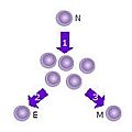
 Clonal Expansion of T Cells.png 1,468 × 1,878; 182 KB
-
 CTL killing strategies.png 3,242 × 2,942; 1.32 MB
-
 Célula T (T Cell) (36461320132).jpg 1,176 × 1,842; 599 KB
-
De-Gedächtnis-T-Zelle.ogg 2.8 s; 28 KB
-
De-Gedächtniszelle.ogg 2.5 s; 24 KB
-
De-T-Zelle.ogg 2.0 s; 19 KB
-
 General Effector Mechanisms of B and T Cells.png 3,300 × 1,650; 554 KB
-
 GZMB cinética y localización.png 1,427 × 774; 718 KB
-
 Helper T Cell Differentiation.png 3,216 × 4,506; 1.64 MB
-
 Human Cell Groups distributed by Cell Count and by Aggregate Cell Mass.jpg 3,162 × 2,096; 1.08 MB
-

-
 Immune Response.svg 512 × 384; 65 KB
-
 Immunocompetencef.png 2,428 × 1,888; 5.83 MB
-
-
 Immunological Memory.png 5,025 × 4,579; 1.55 MB
-
 Immunoregulation by regulatory T cells (Treg)..jpg 432 × 311; 46 KB
-
 Interaccions establertes per LIGHT.jpg 2,338 × 1,700; 281 KB
-
 Lieferkette bei der CAR-T-Zelltherapie.webp 1,347 × 1,058; 65 KB
-
 Linfocitos T de memoria.png 1,280 × 720; 174 KB
-
 Nickel allergy T-cells.jpg 743 × 586; 68 KB
-
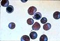
 Normal T Cells (6830348943).jpg 106 × 160; 4 KB
-
 Normal T Cells (6830364101).jpg 160 × 109; 4 KB
-

-

-
-
 Presentación antigénica.png 1,412 × 744; 257 KB
-
 Reconeixement.jpg 497 × 176; 15 KB
-
 Sistèma immunitari - Activacion dei linfocits T dins l'òme.png 1,066 × 1,562; 380 KB
-
 Sistèma immunitari - Activacion dei linfocits T.png 1,079 × 1,582; 363 KB
-
 T Cell (30107399504).jpg 1,633 × 2,256; 469 KB
-
 T cell activation-Deutsch.png 686 × 1,038; 113 KB
-
 T cell activation-es.png 686 × 1,038; 92 KB
-
 T cell activation-eu.png 749 × 885; 131 KB
-
 T cell activation-ja.svg 328 × 496; 41 KB
-
 T cell activation-tr.png 686 × 1,038; 229 KB
-
 T cell activation.png 686 × 1,038; 155 KB
-
 T cell prolif.jpg 150 × 150; 3 KB
-
 T Lymphocyte (16760076824).jpg 2,560 × 1,920; 3.04 MB
-
 T Lymphocyte (16760110354).jpg 640 × 480; 49 KB
-
 T Lymphocyte (17382239521).jpg 2,560 × 1,920; 2.73 MB
-
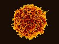
 T Lymphocyte, also known as a T cell (yellow color).jpg 2,428 × 1,888; 3.2 MB
-
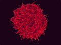
 T Lymphocyte.jpg 2,560 × 1,920; 3.4 MB
-
 Westgate Microscopy t-lymphocytes.jpg 1,113 × 611; 610 KB


.webp)
.png)


_(36461320132).jpg)


..jpg)


.jpg)
.jpg)
.dark.svg)
.svg)
.yellow_Wikidata_Q14904995.svg)


.jpg)


.jpg)
.jpg)
.jpg)
.jpg)

