Category:Tight junctions
Jump to navigation
Jump to search
type of cellular connection that minimizes metabolic flows between adjacent cells  | |||||
| Upload media | |||||
| Instance of |
| ||||
|---|---|---|---|---|---|
| Subclass of |
| ||||
| |||||
Subcategories
This category has the following 6 subcategories, out of 6 total.
Media in category "Tight junctions"
The following 58 files are in this category, out of 58 total.
-
 Aves gastrulation Mechanism of cell ingression.jpg 1,478 × 1,127; 175 KB
-
 Aves gastrulation Origins of flow organisation.jpg 1,660 × 1,579; 661 KB
-
 Cellular tight junction-cz.svg 499 × 646; 145 KB
-
 Cellular tight junction-fr.png 800 × 1,036; 182 KB
-
 Commensals vs pathogens mechanism.png 834 × 768; 223 KB
-
 Commensals vs pathogens mechanism.svg 483 × 445; 144 KB
-
 Commensals vs pathogens mechanisms.png 3,863 × 3,558; 1.35 MB
-
-
 Esquema epitelio intestinal.png 856 × 604; 208 KB
-
 Esquema permeabilidad intestinal aumentada.png 1,004 × 1,215; 113 KB
-
 Figure 04 06 03.jpg 544 × 451; 168 KB
-
 Honeycomb cells-2.jpg 1,376 × 1,032; 529 KB
-
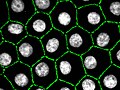 Honeycomb cells.jpg 1,376 × 1,032; 142 KB
-
-
-
-
-
-
 Increased intestinal permeability.png 1,004 × 1,094; 693 KB
-
 Jonction serr.png 513 × 473; 30 KB
-
 Junctional complex and pinocytotic vesicles - embryonic brain-TEM.jpg 1,600 × 1,278; 939 KB
-
 Localització de la proteïna PALS1.jpg 1,944 × 1,073; 120 KB
-
MDCK-Cystogenesis-Driven-by-Cell-Stabilization-within-Computational-Analogues-pcbi.1002030.s019.ogv 1 min 20 s, 550 × 550; 1.46 MB
-
MDCK-Cystogenesis-Driven-by-Cell-Stabilization-within-Computational-Analogues-pcbi.1002030.s020.ogv 1 min 20 s, 550 × 550; 1.54 MB
-
 Membrane proteins of the polarized hepatocytes.jpg 1,039 × 730; 567 KB
-
 Neuro-anatomy 01.png 1,617 × 1,178; 1.87 MB
-
-
Regulation-of-Embryonic-Cell-Adhesion-by-the-Prion-Protein-pbio.1000055.sv001.ogv 8.0 s, 299 × 299; 1.19 MB
-
Regulation-of-Embryonic-Cell-Adhesion-by-the-Prion-Protein-pbio.1000055.sv002.ogv 8.0 s, 356 × 357; 1.66 MB
-
Regulation-of-Embryonic-Cell-Adhesion-by-the-Prion-Protein-pbio.1000055.sv003.ogv 12 s, 158 × 155; 88 KB
-
Regulation-of-Embryonic-Cell-Adhesion-by-the-Prion-Protein-pbio.1000055.sv004.ogv 12 s, 158 × 156; 92 KB
-
Regulation-of-Embryonic-Cell-Adhesion-by-the-Prion-Protein-pbio.1000055.sv005.ogv 2.1 s, 1,024 × 1,024; 1.68 MB
-
Regulation-of-Embryonic-Cell-Adhesion-by-the-Prion-Protein-pbio.1000055.sv006.ogv 12 s, 523 × 512; 340 KB
-
 Selective permeability routes in epithelium.png 1,142 × 821; 856 KB
-
 Selective permeability routes in epithelium.svg 960 × 720; 396 KB
-
 Septatejunction.jpg 369 × 393; 60 KB
-
 The Biological bulletin (20352856166).jpg 2,024 × 2,092; 1.31 MB
-
 The Biological bulletin (20379118525).jpg 1,996 × 2,096; 1.3 MB
-
 Tight junction 03 CA.png 2,461 × 2,004; 1.33 MB
-
 Tight junction 03.png 2,458 × 2,004; 1.33 MB
-
 Tight junction blowup.jpg 882 × 751; 389 KB
-
 Tight Junction Transmembrane Proteins.jpg 1,254 × 1,034; 231 KB
-
 Tight junction.png 177 × 278; 37 KB
-
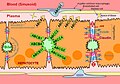
 Tightjunction BBB.jpg 311 × 390; 58 KB
-
 TJschema.png 514 × 488; 28 KB
-
-
-
-
-
Tyrosine-kinase-mediated-axial-motility-of-basal-cells-revealed-by-intravital-imaging-ncomms10666-s2.ogv 7.0 s, 1,163 × 387; 739 KB
-
Tyrosine-kinase-mediated-axial-motility-of-basal-cells-revealed-by-intravital-imaging-ncomms10666-s3.ogv 17 s, 1,190 × 720; 551 KB
-
Tyrosine-kinase-mediated-axial-motility-of-basal-cells-revealed-by-intravital-imaging-ncomms10666-s4.ogv 9.0 s, 1,185 × 714; 1.15 MB
-
-
Tyrosine-kinase-mediated-axial-motility-of-basal-cells-revealed-by-intravital-imaging-ncomms10666-s6.ogv 13 s, 1,024 × 1,024; 4.31 MB
-
Tyrosine-kinase-mediated-axial-motility-of-basal-cells-revealed-by-intravital-imaging-ncomms10666-s7.ogv 21 s, 1,185 × 714; 2.61 MB
-
Tyrosine-kinase-mediated-axial-motility-of-basal-cells-revealed-by-intravital-imaging-ncomms10666-s8.ogv 31 s, 1,050 × 576; 1.54 MB
-
 Unctional complex and pinocytotic vesicles - embryonic brain - TEM.jpg 1,600 × 1,278; 861 KB
-


.jpg)
.jpg)


