Category:Visual cortex
Jump to navigation
Jump to search
region of the brain that processes visual information | |||||
Upload media | |||||
Instance of |
| ||||
---|---|---|---|---|---|
Subclass of |
| ||||
Part of | |||||
| |||||
![]() |
see also
- Category:Auditory cortex
- Category:Somatosensory cortex
- Category:Motor cortex
- Category:Language cortex
and
Subcategories
This category has the following 13 subcategories, out of 13 total.
B
- Brodmann area 18 (16 F)
- Brodmann area 19 (14 F)
C
H
L
- Lunate sulcus (2 F)
M
- Mus musculus visual cortex (13 F)
O
- Occipital pole (6 F)
P
- Pericalcarine cortex (1 F)
R
- Retinotopy (13 F)
V
- Ventral and dorsal stream (16 F)
- Visual area MT (13 F)
- Visual area V4 (18 F)
Media in category "Visual cortex"
The following 128 files are in this category, out of 128 total.
- 1204 Optic Nerve vs Optic Tract fr.jpg 346 × 318; 21 KB
- Agrégat de cartes de champ visuel de Wandell et als 2007.png 472 × 839; 332 KB
- BA17 Primary visual cortex - animation.gif 300 × 300; 1.77 MB
- BA17 Primary visual cortex - lateral view.png 1,200 × 1,200; 689 KB
- BA17 Primary visual cortex - medial view.png 1,200 × 1,200; 532 KB
- BA17 Primary visual cortex - posterior view.png 1,200 × 1,200; 436 KB
- BA17,18,19 - Visual cortex (V1, V2, V3) - animation.gif 300 × 300; 1.62 MB
- BA17,18,19 - Visual cortex (V1, V2, V3) - lateral view.png 1,200 × 1,200; 693 KB
- BA17,18,19 - Visual cortex (V1, V2, V3) - medial view.png 1,200 × 1,200; 533 KB
- BA17,18,19 - Visual cortex (V1, V2, V3) - posterior view.png 1,200 × 1,200; 441 KB
- BA18 - Secondary visual cortex (V2) - animation.gif 300 × 300; 1.73 MB
- BA18 - Secondary visual cortex (V2) - lateral view.png 1,200 × 1,200; 689 KB
- BA18 - Secondary visual cortex (V2) - medial view.png 1,200 × 1,200; 533 KB
- BA18 - Secondary visual cortex (V2) - posterior view.png 1,200 × 1,200; 438 KB
- BA19 - Visual association cortex (V3) - animation.gif 300 × 300; 1.72 MB
- BA19 - Visual association cortex (V3) - lateral view.png 1,200 × 1,200; 686 KB
- BA19 - Visual association cortex (V3) - medial view.png 1,200 × 1,200; 531 KB
- BA19 - Visual association cortex (V3) - posterior view.png 1,200 × 1,200; 435 KB
- Blausen 0103 Brain Sensory&Motor ar.png 1,600 × 960; 5.87 MB
- Blausen 0103 Brain Sensory&Motor.png 1,600 × 960; 993 KB
- Brain 2.jpg 721 × 540; 171 KB
- Brain image of blind echolocator.tif 1,200 × 923; 882 KB
- Brodmann areas 17 18 19.png 256 × 192; 40 KB
- Brodmann Cytoarchitectonics 17.png 599 × 952; 231 KB
- Brodmann Cytoarchitectonics 18.png 599 × 952; 225 KB
- Brodmann Cytoarchitectonics 19.png 599 × 952; 247 KB
- Cajal cortex drawings.png 672 × 777; 181 KB
- Candelabro 03 CUATRIS.jpg 377 × 445; 128 KB
- Carte blobs.png 316 × 302; 68 KB
- Carte retino cluster.png 886 × 531; 357 KB
- Caudal view of a brain after dissection.png 1,890 × 2,405; 2.94 MB
- Cerebral hemisphere of an ape. Wellcome L0001994.jpg 1,670 × 1,122; 698 KB
- Cerebro sistema.png 472 × 314; 57 KB
- Complex cell.JPG 214 × 196; 5 KB
- Constudeyepath ja.png 800 × 1,139; 112 KB
- Constudeyepath1.JPG 975 × 1,265; 441 KB
- Constudproc.png 763 × 865; 31 KB
- Cortex strie.svg 305 × 393; 39 KB
- Corticalorient.GIF 450 × 327; 5 KB
- Coverage-continuity-and-visual-cortical-architecture-2042-1001-1-17-S1.ogv 22 s, 1,024 × 418; 3.21 MB
- Coverage-continuity-and-visual-cortical-architecture-2042-1001-1-17-S2.ogv 22 s, 1,024 × 418; 2.2 MB
- Coverage-continuity-and-visual-cortical-architecture-2042-1001-1-17-S3.ogv 22 s, 1,024 × 418; 3.61 MB
- Coverage-continuity-and-visual-cortical-architecture-2042-1001-1-17-S4.ogv 22 s, 1,024 × 418; 4.06 MB
- Cytoarchitecture Visual cortex.jpg 709 × 809; 212 KB
- Dorsal and ventral attention systems.jpg 878 × 498; 106 KB
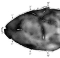
- Ferret brain.jpg 850 × 505; 137 KB
- Functional magnetic resonance imaging.jpg 250 × 208; 11 KB
- Fusiform face area face recognition.jpg 475 × 503; 95 KB
- Gray726 parieto-occipital sulcus.png 700 × 405; 86 KB
- Gray727 calcarine sulcus.svg 1,025 × 598; 22 KB
- Gray727 Cuneus.png 1,025 × 598; 144 KB
- Gray727 parieto-occipital fissure.svg 1,025 × 598; 22 KB
- Gray746.png 329 × 650; 21 KB
- Gray756.png 600 × 354; 32 KB
- Gray757.png 600 × 346; 27 KB
- Human visual cortex V1 cropped.png 900 × 349; 331 KB
- Human visual cortex V1.png 901 × 961; 820 KB
- Hyperbolic-Planforms-in-Relation-to-Visual-Edges-and-Textures-Perception-pcbi.1000625.s006.ogv 1.5 s, 434 × 343; 364 KB
- Hypercolonne.png 927 × 596; 88 KB
- Hypercolonne.svg 294 × 230; 24 KB
- ImplantSawan.JPG 512 × 384; 15 KB
- Latency of response to a visual stimulus along the visual pathways.jpg 1,999 × 1,504; 393 KB
- Lateral and medial views of the marmoset cerebral cortex.jpg 2,667 × 793; 152 KB
- Lawrence 1960 9.3.png 1,784 × 2,792; 353 KB
- Lisa analysis.png 404 × 556; 136 KB
- Location and visuotopic organization of marmoset primary visual cortex (V1).jpg 1,733 × 1,717; 172 KB
- Long-Evans rat V1 cortico-connection experiment - Fluorescent axon bundles in left V1.jpg 4,288 × 2,848; 4.56 MB
- Low-Level-Contrast-Statistics-of-Natural-Images-Can-Modulate-the-Frequency-of-Event-Related-Video1.ogv 35 s, 1,280 × 960; 4.02 MB
- Low-Level-Contrast-Statistics-of-Natural-Images-Can-Modulate-the-Frequency-of-Event-Related-Video2.ogv 35 s, 1,280 × 960; 3.54 MB
- Mapping-Human-Whole-Brain-Structural-Networks-with-Diffusion-MRI-pone.0000597.s007.ogv 6.8 s, 1,008 × 1,008; 2.86 MB
- Medial surface of cerebral cortex - ceneus.png 1,179 × 747; 514 KB
- Microglial-P2Y12-is-necessary-for-synaptic-plasticity-in-mouse-visual-cortex-ncomms10905-s2.ogv 3.0 s, 379 × 391; 138 KB
- Mod hierarchique Vasseur.svg 283 × 393; 26 KB
- Module.PNG 702 × 539; 34 KB
- Module2.PNG 702 × 539; 35 KB
- Module3.PNG 702 × 539; 37 KB
- Neocórtex visual capas.png 305 × 600; 917 KB
- Neural Abstraction Pyramid.jpg 1,343 × 782; 253 KB
- Optic nerve pair & two brain hemispheres.jpg 252 × 405; 21 KB
- Optic pathway.png 1,890 × 2,528; 1.29 MB
- Optical-transformations.png 1,415 × 2,085; 411 KB
- Orientation V1.svg 354 × 283; 49 KB
- Parcellation of different cortical regions involved in visual processing.jpg 1,800 × 1,079; 205 KB
- Proj retine CGL cortex colonnes1.jpg 1,546 × 2,709; 326 KB
- Proj retine CGL cortex1.svg 549 × 857; 134 KB
- Schematic organization of visual cortex in the marmoset.jpg 2,667 × 2,289; 355 KB
- Scissure calca1.svg 354 × 461; 45 KB
- Sehbahn 3D v2.jpg 817 × 674; 94 KB
- Sehbahn 3D.jpg 817 × 674; 99 KB
- Sensitivity of human V6 to Translational egomotion.jpg 744 × 603; 231 KB
- Surface models and borders visual area.jpg 680 × 1,033; 372 KB
- Unsupervised-Learning-of-Visual-Features-through-Spike-Timing-Dependent-Plasticity-pcbi.0030031.sv001.ogv 2 min 22 s, 470 × 490; 4.73 MB
- Unsupervised-Learning-of-Visual-Features-through-Spike-Timing-Dependent-Plasticity-pcbi.0030031.sv002.ogv 3 min 20 s, 470 × 490; 8.67 MB
- Unsupervised-Learning-of-Visual-Features-through-Spike-Timing-Dependent-Plasticity-pcbi.0030031.sv003.ogv 5 min 0 s, 470 × 490; 10.3 MB
- V1 v2 v3.jpg 522 × 460; 32 KB
- Visual analisation.jpg 311 × 542; 48 KB
- Visual cortex - intermed mag.jpg 2,848 × 4,272; 8.74 MB
- Visual cortex - low mag.jpg 2,848 × 4,272; 5.43 MB
- Visual cortex near fracture.png 819 × 784; 448 KB
- Visual cortex.jpg 589 × 432; 37 KB
- Visual field maps.jpg 886 × 886; 106 KB
- Visualcortex.gif 201 × 179; 10 KB
- VisualSingularity.jpg 599 × 393; 55 KB
- Visueel systeem.PNG 777 × 368; 17 KB
- Visueller Cortex(MRT).gif 393 × 320; 69 KB
- Voies visuelles3.svg 744 × 964; 151 KB
- Voxel-man-brain.jpg 900 × 900; 497 KB
- Wandell maps.jpg 1,181 × 886; 114 KB
- Wat waar.PNG 616 × 443; 113 KB
- Ієрархія кіркової репрезентації.png 1,731 × 946; 1.07 MB
- Зоровий шлях.jpg 250 × 218; 45 KB