Commons:Wiki Science Competition 2023/Image sets
Jump to navigation
Jump to search

Argentina · Estonia · Finland · France and Monaco · Indonesia · Ireland · Italy · Malaysia · Nigeria · North Macedonia · Poland · Russia · South Africa · Spain · Switzerland · Ukraine · United States ··· The rest of the World
People in Science · Microscopy images · Non-photographic media · Wildlife & nature · Astronomy images · General category · Image sets ··· Winners
Wiki Science Competition 2023 had 818 files submitted by 94 uploaders in the image sets category.
Here are the finalists.
-
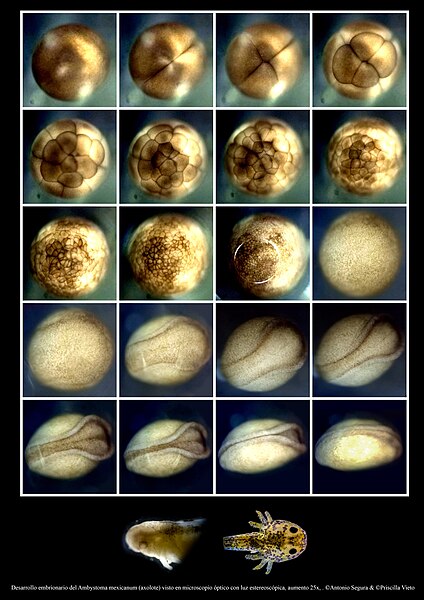 Development of Ambystoma mexicanum axolotl, the process took a total of 18 days, showcasing all phases including gastrulation, morula, and neurulation. Method: stereoscopic microscopy. Photos by Brandon Antonio Segura Torres & Priscilla Vieto Bonilla

-
 Close-up of the compound eye of Drosophila melanogaster (fruit fly) observed under a scanning electron microscope, digitally colored. Photo by Brandon Antonio Segura Torres & Priscilla Vieto Bonilla
.jpg)
-
 Mite from the Pheroliodidae family, observed under a scanning electron microscope. Magnification 181x. What appears to be 'wings' is actually a pseudo-stigmatic organ. Photos by Brandon Antonio Segura Torres & Priscilla Vieto Bonilla

-
 Didymo algae (Didymosphenia geminata) observed under a scanning electron microscope. In the first image, Didymo (the larger one) is seen in valve view surrounded by smaller diatoms. Subsequently, there is an increase in magnification to focus on the raphe and its ornamentations. Finally, the diatom is observed in girdle view. Photos by Brandon Antonio Segura Torres & Priscilla Vieto Bonilla

-
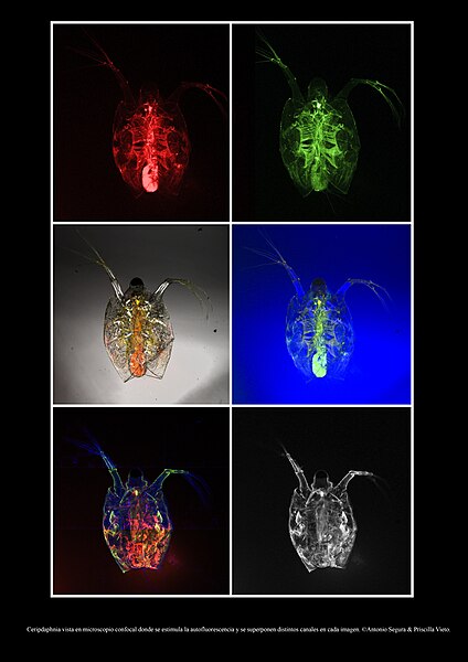 Specimen of Daphnia with autofluorescence observed under a confocal microscope in different channels. This specimen was not stained, allowing the natural fluorescence from its interior, which includes chlorophyll from its diet, to be visualized. Photos by Brandon Antonio Segura Torres & Priscilla Vieto Bonilla

-
 Filamentous microalgae viewed under four different lighting conditions using an immersion optical microscope: bright field, dark field, polarized light, and polarized light with dark field, respectively. Photos by Brandon Antonio Segura Torres & Priscilla Vieto Bonilla

-
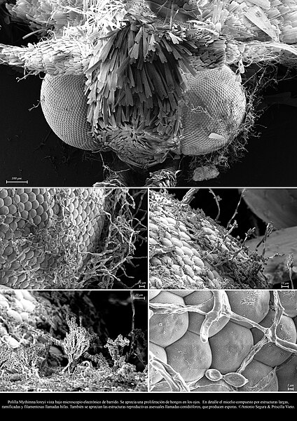 Moth of the species Mythimna loreyi with proliferation of Penicillium fungi in its eyes, observed under a scanning electron microscope. Photos by Brandon Antonio Segura Torres & Priscilla Vieto Bonilla

-

-

-

-

-

-
 Organisms found on water. Photos by Janek Lass


.jpg)


-
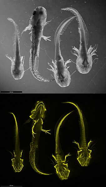 Analysis of collagen expression in young salamanders via HCR FISH visualization. Photos by Lennart Rikk

-

-
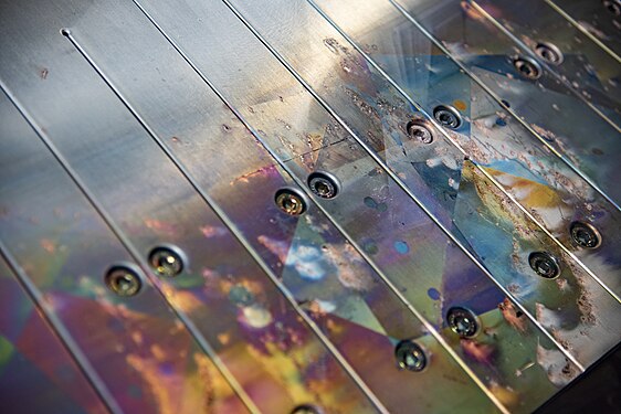
 Investigating the composition of iron. Photos by Kristo Oks
 -


-

-
-

-
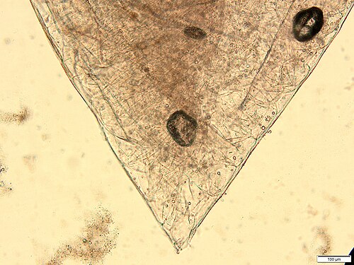
 Reference collection work for archaeological dental calculus. Photos by Agnes Unt


-

-

-

-

-
 Tunnels, samples and equipment in Onkalo spent nuclear fuel repository. Photos by Kallerna


-

-

-

-

-
-

-

-
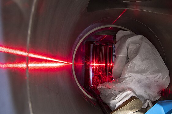
 WEST tokamak. Cadarache, France. Photos by Hélène Roche et Christophe Roux / CEA IRFM


-

-

-

-

-

-

-
-
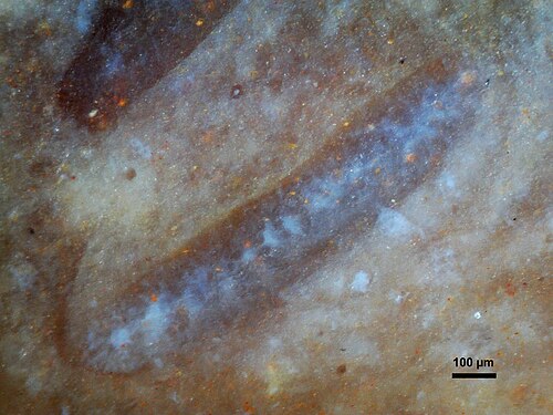
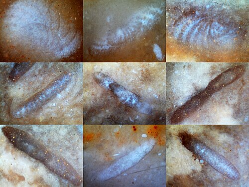 Choffatella decipiens Schlumberger Em. Maync, 1950. Stage : Lower Cretaceous, Barremian - Aptian, Urgonian Facies. Location : South-East France. Photos by Benoit Potin
)_1.jpg)
)_2.jpg)
)_3.jpg)
)_4.jpg)
)_5.jpg)
)_6.jpg)
)_7.jpg)
)_8.jpg)
-

-
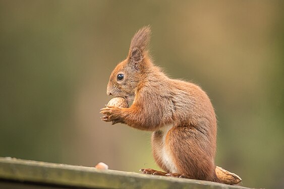 Red squirrel (Sciurus vulgaris). Photos by Jg44.89
 -


-

-

-

-

-
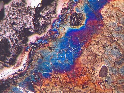
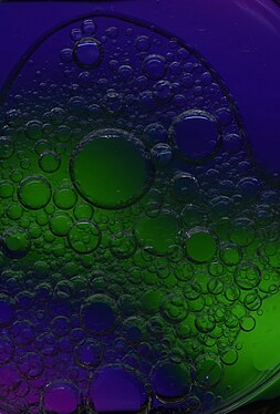 Emulsion of oil and water caused by differences in their chemical and physical properties. Photos by Сибиновска Ангела


