File:A-Drosophila-Model-for-EGFR-Ras-and-PI3K-Dependent-Human-Glioma-pgen.1000374.s017.ogv
Jump to navigation
Jump to search
Size of this JPG preview of this OGG file: 470 × 600 pixels. Other resolutions: 188 × 240 pixels | 376 × 480 pixels | 760 × 970 pixels.
Original file (Ogg Theora video file, length 2.4 s, 760 × 970 pixels, 5.25 Mbps, file size: 1.51 MB)
Captions
Captions
Add a one-line explanation of what this file represents
Summary
[edit]| Description |
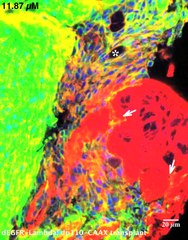
English: An animated 27 µm thick confocal z-stack of dEGFRλ; dp110CAAX tumor cells derived from transplanted larval glia, pictured in Figure 3F. The depth of each frame is noted in the upper left-hand corner. 20 µm scale bar for x/y-axis. Transplanted mutant glia are labeled with membrane bound CD8GFP (green) and the Repo nuclear protein (blue). Actin staining (red, phalloidin) reveals abdominal anatomy of the host. Arrowheads in indicate dEGFRλ;dp110CAAX glial cells invading an ovary, distinguished by its characteristic actin staining (bright red). An asterisk indicates trachea embedded in this dEGFRλ;dp110CAAX tumor, visible as a hollow actin-positive (red) tubule running through the tissue. Genotypes: Hosts were w1118 virgin females. Transplanted glia were UAS-dEGFRλ UAS-dp110CAAX/+; UAS-CD8GFP/+; repo-Gal4/+. |
||
| Date | |||
| Source | Video S2 from Read R, Cavenee W, Furnari F, Thomas J (2009). "A Drosophila Model for EGFR-Ras and PI3K-Dependent Human Glioma". PLOS Genetics. DOI:10.1371/journal.pgen.1000374. PMID 19214224. PMC: 2636203. | ||
| Author | Read R, Cavenee W, Furnari F, Thomas J | ||
| Permission (Reusing this file) |
|
||
| Provenance |
|
File history
Click on a date/time to view the file as it appeared at that time.
| Date/Time | Thumbnail | Dimensions | User | Comment | |
|---|---|---|---|---|---|
| current | 19:57, 16 November 2012 | 2.4 s, 760 × 970 (1.51 MB) | Open Access Media Importer Bot (talk | contribs) | Automatically uploaded media file from Open Access source. Please report problems or suggestions here. |
You cannot overwrite this file.
File usage on Commons
There are no pages that use this file.
Transcode status
Update transcode statusMetadata
Categories:
- Videos of cell signaling
- Cancer genetics
- Videos of disease models
- Videos of cell cycle
- Cell cycle proteins
- Dysplasias
- Animal models of disease
- Videos of Drosophila melanogaster proteins
- Videos of gliomas
- Neuroglia
- Phosphatidylinositol 3-kinases
- Epidermal growth factor receptor
- Videos of signal transduction
- Ras proteins
- Genotypes