File:Adenomyosis, Hysterectomy Specimen (640264025).jpg
Jump to navigation
Jump to search

Size of this preview: 800 × 476 pixels. Other resolutions: 320 × 191 pixels | 640 × 381 pixels | 1,024 × 610 pixels | 1,280 × 762 pixels | 2,560 × 1,524 pixels | 4,229 × 2,518 pixels.
Original file (4,229 × 2,518 pixels, file size: 3.01 MB, MIME type: image/jpeg)
Captions
Captions
Add a one-line explanation of what this file represents
Summary[edit]
.jpg&action=edit§ion=1)
| Description |
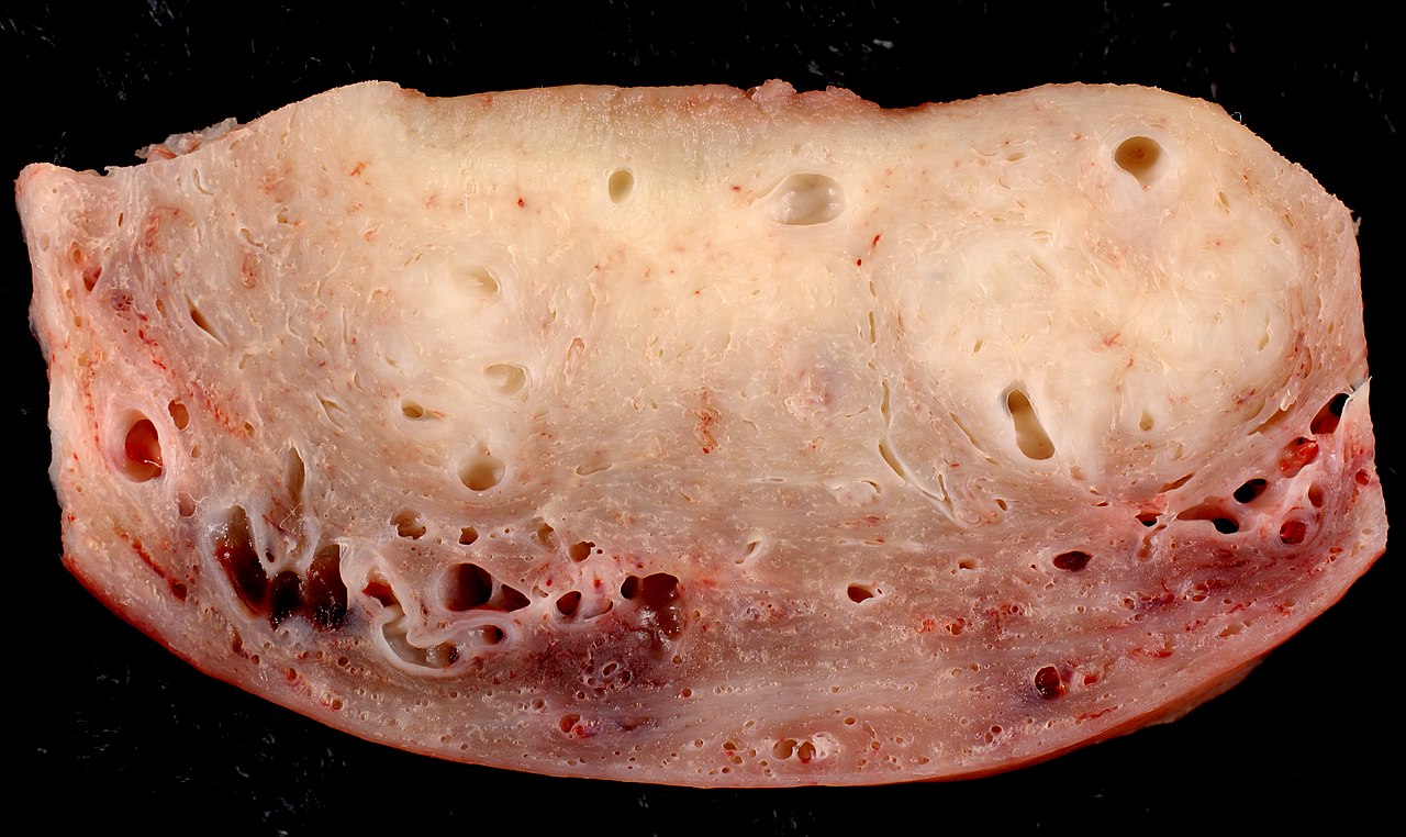
Cross section through the wall of a hysterectomy specimen of a 30-year-old woman who reported chronic pelvic pain and abnormal uterine bleeding. The endometrial surface is at the top of the image, and the serosa is at the bottom. I think most cases of adenomyosis can be reliably diagnosed grossly by an experienced prosector examining a fixed specimen. Following formalin fixation, the soft adenomyotic areas stand out more strikingly against the firmer myometrium. |
| Date | |
| Source | Adenomyosis, Hysterectomy Specimen |
| Author | Ed Uthman from Houston, TX, USA |
Licensing[edit]
.jpg&action=edit§ion=2)
This file is licensed under the Creative Commons Attribution 2.0 Generic license.
- You are free:
- to share – to copy, distribute and transmit the work
- to remix – to adapt the work
- Under the following conditions:
- attribution – You must give appropriate credit, provide a link to the license, and indicate if changes were made. You may do so in any reasonable manner, but not in any way that suggests the licensor endorses you or your use.
| This image was originally posted to Flickr by euthman at https://flickr.com/photos/78147607@N00/640264025. It was reviewed on 29 October 2020 by FlickreviewR 2 and was confirmed to be licensed under the terms of the cc-by-2.0. |
File history
Click on a date/time to view the file as it appeared at that time.
| Date/Time | Thumbnail | Dimensions | User | Comment | |
|---|---|---|---|---|---|
| current | 09:26, 29 October 2020 | | 4,229 × 2,518 (3.01 MB) | Netha Hussain (talk | contribs) | Transferred from Flickr via #flickr2commons |
You cannot overwrite this file.
File usage on Commons
There are no pages that use this file.
.jpg&oldid=797894610)