File:Birefringence microscopy of pseudogout, annotated.jpg
跳转到导航
跳转到搜索
本预览的尺寸:662 × 600像素。 其他分辨率:265 × 240像素 | 530 × 480像素 | 848 × 768像素 | 1,130 × 1,024像素 | 1,745 × 1,581像素。
原始文件 (1,745 × 1,581像素,文件大小:615 KB,MIME类型:image/jpeg)
说明
说明
添加一行文字以描述该文件所表现的内容
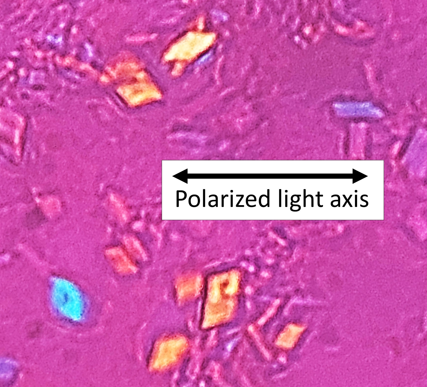
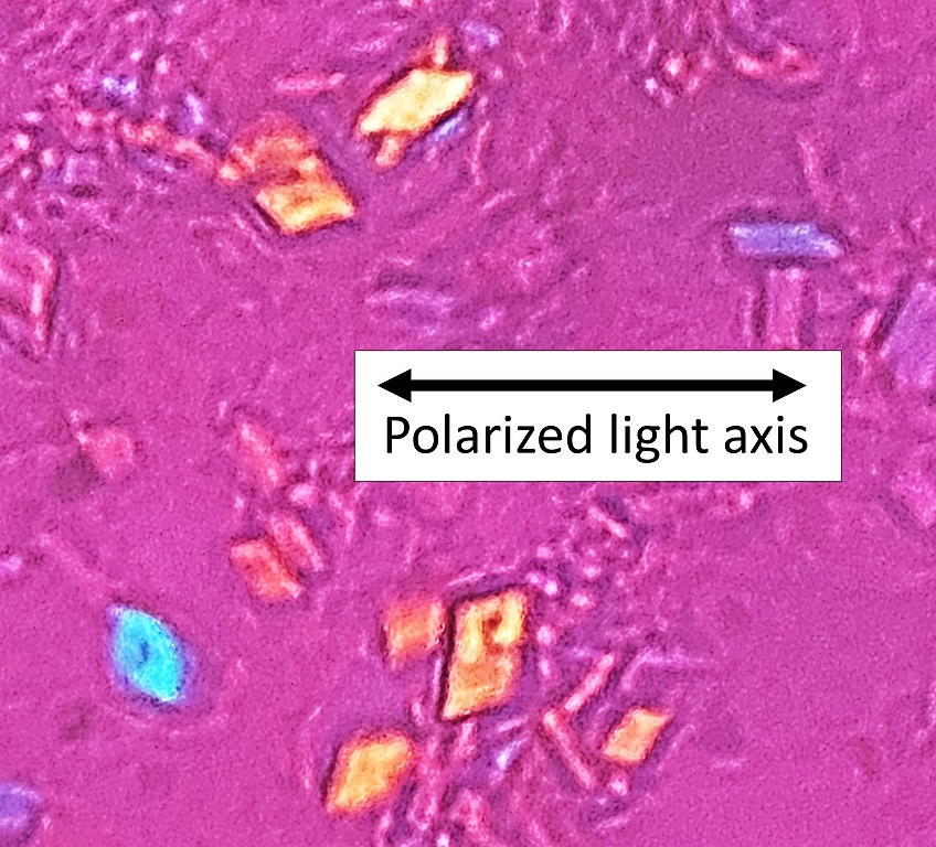
Birefringence microscopy of pseudogout, annotated
摘要
[编辑]
| 描述 |
English: Microscopy with polarized light of tissue by a metatarsal joint, showing crystals whereof some (one annotated) have rhomboid shape and weak positive nirefringence, consistent with calcium pyrophosphate dihydrate crystal deposition disease (pseudogout). |
| 日期 | |
| 来源 | 自己的作品 |
| 作者 |
.jpg) - Reusing images - Conflicts of interest: None Consent note: Consent from the patient or patient's relatives is regarded as redundant, because of absence of identifiable features (List of HIPAA identifiers) in the media and case information (See also HIPAA case reports guidance). |
| 其他版本 |
 |
| 相机位置 | | 在以下服务上查看本图像和附近其他图像: OpenStreetMap |
|---|
许可协议
[编辑]
| 本作品采用知识共享CC0 1.0 通用公有领域贡献许可协议授权。 | |
| 采用本宣告发表本作品的人,已在法律允许的范围内,通过在全世界放弃其对本作品拥有的著作权法规定的所有权利(包括所有相关权利),将本作品贡献至公有领域。您可以复制、修改、传播和表演本作品,将其用于商业目的,无需要求授权。
|
文件历史
点击某个日期/时间查看对应时刻的文件。
| 日期/时间 | 缩略图 | 大小 | 用户 | 备注 | |
|---|---|---|---|---|---|
| 当前 | 2022年4月4日 (一) 22:59 | | 1,745 × 1,581(615 KB) | Mikael Häggström(留言 | 贡献) | Sharper |
| 2020年11月12日 (四) 14:45 |  | 628 × 567(82 KB) | Mikael Häggström(留言 | 贡献) | +Axis | |
| 2020年11月12日 (四) 14:39 |  | 473 × 426(48 KB) | Mikael Häggström(留言 | 贡献) | Uploaded a work by {{Mikael Häggström|cat=Micrographs|consent=noid}} from {{Own}} with UploadWizard |
您不可以覆盖此文件。
文件用途
全域文件用途
以下其他wiki使用此文件:
- ar.wikipedia.org上的用途
- en.wikipedia.org上的用途
- zh.wikipedia.org上的用途