File:Bronchial anatomy.jpg
Qallariy willañiqi (2646 × 2048 iñu; willañiqip chhikan kaynin: 1,98 MB; MIME laya: image/jpeg)
Leyendas
Leyendas
Pisichay[llamk'apuy]
| T'iktuna |
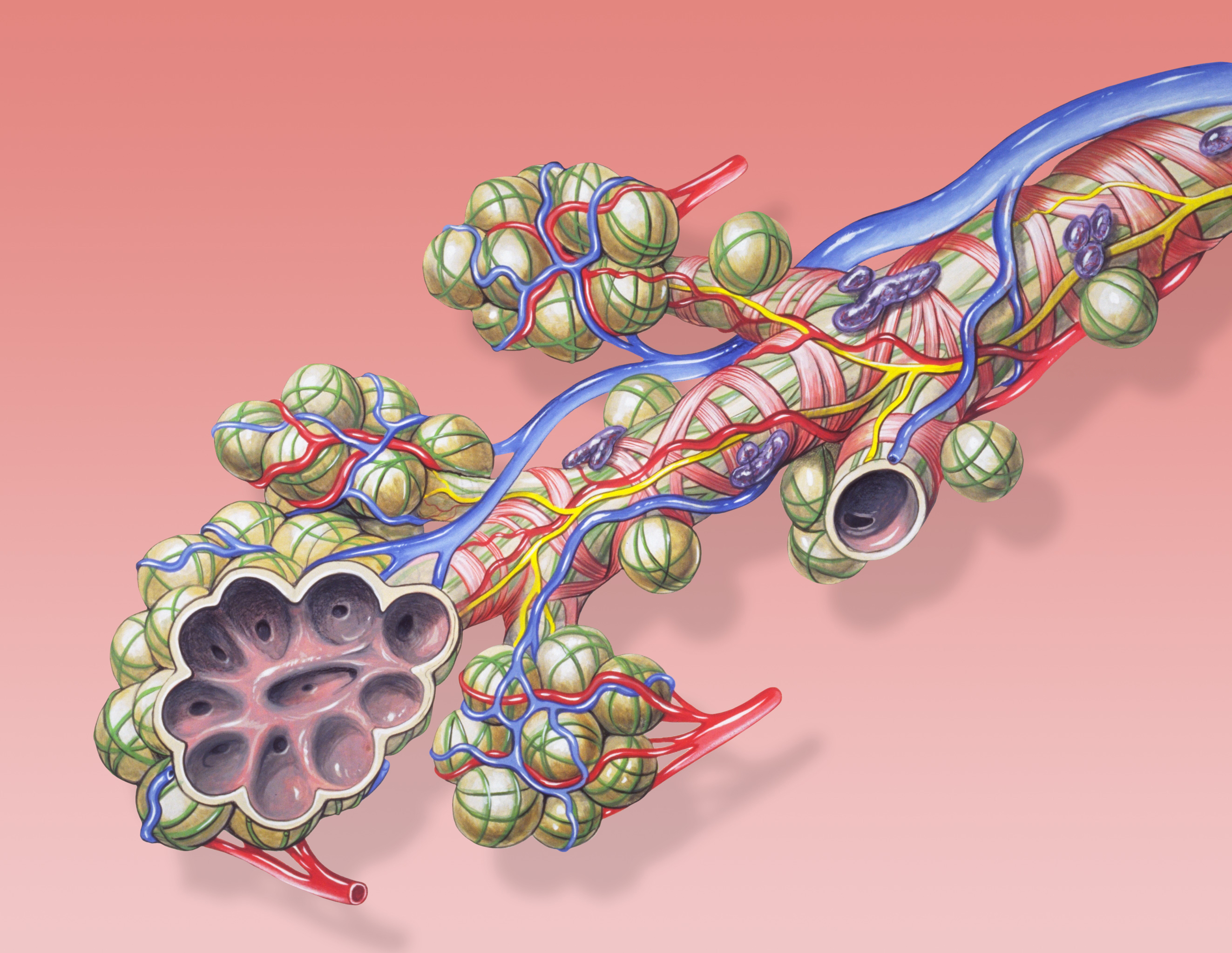
English: Bronchial anatomy detail of alveoli and lung circulation.
Français : Anatomie pulmonaire: détail des alvéoles et de la circulation pulmonaires . |
| P'unchaw | |
| Pukyu | Patrick J. Lynch, medical illustrator |
| Ruraq | Patrick J. Lynch, medical illustrator |
| Saqillay (Kay willañiqita musuqmanta llamk'achiy) |
Creative Commons Attribution 2.5 License 2006 |
| Huk musuqchasqakuna | Derivative works of this file: Bronchial anatomy Cerchiato.png None |
|

This image was selected as picture of the day on Wikimedia Commons for 16 January 2012. It was captioned as follows: English: Bronchial anatomy detail of alveoli and lung circulation. Other languages:
English: Bronchial anatomy detail of alveoli and lung circulation. Español: Anatomía bronquial: detalle de los alvéolos y la circulación pulmonar. Français : Anatomie pulmonaire : détail des alvéoles et de la circulation pulmonaires. Italiano: L'anatomia delle ramificazioni terminali dell'albero respiratorio con l'annessa vascolarizzazione (arteria e vene polmonari). Nederlands: Anatomisch detail van longblaasjes (pulmonaire alveoli) in de longen, waar tijdens de ademhaling de gaswisseling plaatsgrijpt. Русский: Анатомия бронха Українська: Анатомія бронха з розрізом альвеол, бронхіальні частини легеневої артерії і легеневої вени, спинна частина легеневої гілки блукаючого нерва. ქართული: ბრონქების ანატომია დეტალურად. 日本語: 肺胞と肺循環を図解する気管支の解剖図。 中文: 肺泡解刨细节和肺循环。 |
Patrick J. Lynch; illustrator; C. Carl Jaffe; MD; cardiologist Yale University Center for Advanced Instructional Media Medical Illustrations by Patrick Lynch, generated for multimedia teaching projects by the Yale University School of Medicine, Center for Advanced Instructional Media, 1987-2000. Patrick J. Lynch, http://patricklynch.net Creative Commons Attribution 2.5 License 2006; no usage restrictions except please preserve our creative credits: Patrick J. Lynch, medical illustrator; C. Carl Jaffe, MD, cardiologist. https://creativecommons.org/licenses/by/2.5/
Saqillaspa[llamk'apuy]
- Qispilla rurarillay:
- rakinakuy – iskaychay, mast'ariy, maymanpas kachay kay rurasqata
- musuqmanta chaqruy – rurasqata tupayay
- Kay phatakuna tukuptinqa:
- Ruraqpa sutinta willay – Kay rurasqataqa ruraqninpa icha saqillaqninpa sut'ichasqan hina unanchanaykim (ichataq amapuni kay hinachu, pay q'imisunkiman icha rurayniykita q'iminman rikch'akunman).
| Annotations | This image is annotated: View the annotations at Commons |
Willañiqip wiñay kawsaynin
P'unchaw/pacha nisqapi ñit'iy chaypacha willañiqi kachkasqata qhawanaykipaq.
| P'unchaw/Pacha | Uchuylla rikchacha | Chhikanyachikuqkuna | Ruraq | Willapuy | |
|---|---|---|---|---|---|
| kunan | 11:31 4 awu 2010 | | 2646 × 2048 (1,98 MB) | Dcoetzee (rimanakuy | llamk'apusqakuna) | Remove watermark |
| 04:49 26 dis 2006 |  | 2646 × 2048 (1,42 MB) | Patrick.lynch (rimanakuy | llamk'apusqakuna) | {{Information |Description = Bronchial anatomy detail of alveoli and lung circulation |Source = Patrick J. Lynch, medical illustrator |Date = December 23, 2006 |Author = Patrick J. Lynch, medical illustrator |Permission = Creative Commons Attribution 2.5 |
Manam atinkichu kay willañiqita huknachayta.
Maypim willañiqita llamk'achinku
Kay rikchamanqa kay qatiq 40 p'anqakunam t'inkimun:
- User:Miya/POTD
- User:Ö/Best/2010
- User talk:99of9/Promotions
- Commons:Featured picture candidates/File:Bronchial anatomy.jpg
- Commons:Featured picture candidates/Log/August 2010
- Commons:Featured pictures/Non-photographic media/Computer-generated
- Commons:Featured pictures/chronological/2010-B
- Commons:Picture of the Year/2010/Galleries/Diagrams
- Commons:Picture of the Year/2010/Galleries/Diagrams/Large
- Commons:Picture of the Year/2010/Galleries/Diagrams/Small
- Commons:Picture of the Year/2010/Galleries/Index/9
- Commons:Picture of the Year/2010/Galleries/Index/Diagrams
- Commons:Picture of the Year/2010/Galleries/Table
- Commons:Picture of the Year/2010/Galleries/Table/08
- Commons:Picture of the Year/2010/R1/File:Bronchial anatomy.jpg
- Commons:Picture of the Year/2010/Results/R1/ALL/Table
- Commons:Picture of the Year/2010/Results/R1/Category winners
- Commons:Picture of the Year/2010/Results/R1/Checking
- Commons:Picture of the Year/2010/Results/R1/Diagrams
- Commons:Picture of the Year/2010/Results/R1/Diagrams/Table
- Commons talk:Picture of the Year/2010/Galleries/Table
- Commons talk:Picture of the Year/2010/Results/R1/ALL/Table
- File:Bronchial anatomy Cerchiato.png
- Template:Potd/2012-01
- Template:Potd/2012-01-16
- Template:Potd/2012-01-16 (da)
- Template:Potd/2012-01-16 (de)
- Template:Potd/2012-01-16 (en)
- Template:Potd/2012-01-16 (es)
- Template:Potd/2012-01-16 (fr)
- Template:Potd/2012-01-16 (it)
- Template:Potd/2012-01-16 (ja)
- Template:Potd/2012-01-16 (ka)
- Template:Potd/2012-01-16 (ko)
- Template:Potd/2012-01-16 (mk)
- Template:Potd/2012-01-16 (nl)
- Template:Potd/2012-01-16 (ru)
- Template:Potd/2012-01-16 (uk)
- Template:Potd/2012-01-16 (zh-hans)
- Template:Potd/2012-01 (zh-hans)
Mayqin wikikunapi willañiqita llamk'achinku
Kay wakin wikikunam willañiqitaqa llamk'achinku:
- als.wikipedia.org-pi kaykunapi llamk'achinku
- ar.wikipedia.org-pi kaykunapi llamk'achinku
- az.wikipedia.org-pi kaykunapi llamk'achinku
- ba.wikipedia.org-pi kaykunapi llamk'achinku
- be-tarask.wikipedia.org-pi kaykunapi llamk'achinku
- be.wikipedia.org-pi kaykunapi llamk'achinku
- bg.wikipedia.org-pi kaykunapi llamk'achinku
- bn.wikipedia.org-pi kaykunapi llamk'achinku
- bs.wikipedia.org-pi kaykunapi llamk'achinku
- ckb.wikipedia.org-pi kaykunapi llamk'achinku
- crh.wikipedia.org-pi kaykunapi llamk'achinku
- cs.wikipedia.org-pi kaykunapi llamk'achinku
- cv.wikipedia.org-pi kaykunapi llamk'achinku
- da.wikipedia.org-pi kaykunapi llamk'achinku
- de.wikipedia.org-pi kaykunapi llamk'achinku
- de.wikibooks.org-pi kaykunapi llamk'achinku
- en.wikipedia.org-pi kaykunapi llamk'achinku
- en.wikibooks.org-pi kaykunapi llamk'achinku
- en.wikiversity.org-pi kaykunapi llamk'achinku
- eo.wikipedia.org-pi kaykunapi llamk'achinku
- es.wikibooks.org-pi kaykunapi llamk'achinku
- eu.wikipedia.org-pi kaykunapi llamk'achinku
- fa.wikipedia.org-pi kaykunapi llamk'achinku
- fi.wikipedia.org-pi kaykunapi llamk'achinku
- fr.wikipedia.org-pi kaykunapi llamk'achinku
- gl.wikipedia.org-pi kaykunapi llamk'achinku
Qhaway mayqin wikikunapim willañiqita llamk'achinku.