File:Bronchial anatomy.jpg
跳转到导航
跳转到搜索
本预览的尺寸:775 × 600像素。 其他分辨率:310 × 240像素 | 620 × 480像素 | 992 × 768像素 | 1,280 × 991像素 | 2,560 × 1,981像素 | 2,646 × 2,048像素。
原始文件 (2,646 × 2,048像素,文件大小:1.98 MB,MIME类型:image/jpeg)
说明
说明
添加一行文字以描述该文件所表现的内容
摘要
[编辑]| 描述 |
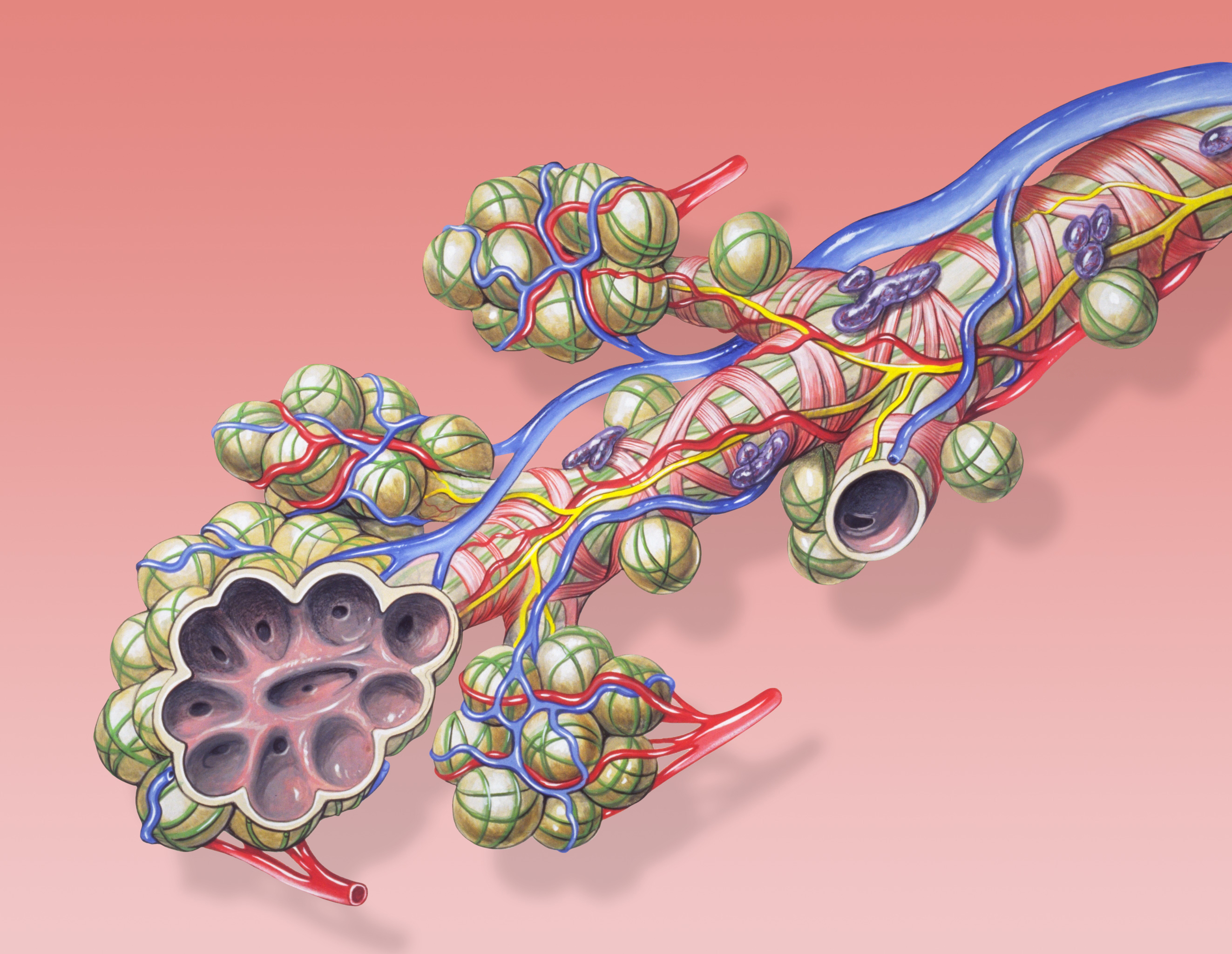
English: Bronchial anatomy detail of alveoli and lung circulation.
Français : Anatomie pulmonaire: détail des alvéoles et de la circulation pulmonaires . |
| 日期 | |
| 来源 | Patrick J. Lynch, medical illustrator |
| 作者 | Patrick J. Lynch, medical illustrator |
| 授权 (二次使用本文件) |
Creative Commons Attribution 2.5 License 2006 |
| 其他版本 | Derivative works of this file: Bronchial anatomy Cerchiato.png None |
|

This image was selected as picture of the day on Wikimedia Commons for 16 January 2012. It was captioned as follows: English: Bronchial anatomy detail of alveoli and lung circulation. Other languages:
English: Bronchial anatomy detail of alveoli and lung circulation. Español: Anatomía bronquial: detalle de los alvéolos y la circulación pulmonar. Français : Anatomie pulmonaire : détail des alvéoles et de la circulation pulmonaires. Italiano: L'anatomia delle ramificazioni terminali dell'albero respiratorio con l'annessa vascolarizzazione (arteria e vene polmonari). Nederlands: Anatomisch detail van longblaasjes (pulmonaire alveoli) in de longen, waar tijdens de ademhaling de gaswisseling plaatsgrijpt. Русский: Анатомия бронха Українська: Анатомія бронха з розрізом альвеол, бронхіальні частини легеневої артерії і легеневої вени, спинна частина легеневої гілки блукаючого нерва. ქართული: ბრონქების ანატომია დეტალურად. 日本語: 肺胞と肺循環を図解する気管支の解剖図。 中文: 肺泡解刨细节和肺循环。 |
Patrick J. Lynch; illustrator; C. Carl Jaffe; MD; cardiologist Yale University Center for Advanced Instructional Media Medical Illustrations by Patrick Lynch, generated for multimedia teaching projects by the Yale University School of Medicine, Center for Advanced Instructional Media, 1987-2000. Patrick J. Lynch, http://patricklynch.net Creative Commons Attribution 2.5 License 2006; no usage restrictions except please preserve our creative credits: Patrick J. Lynch, medical illustrator; C. Carl Jaffe, MD, cardiologist. https://creativecommons.org/licenses/by/2.5/
许可协议
[编辑]
| 注解 | 该图片含有注解:在维基媒体共享资源上查看注解 |
文件历史
点击某个日期/时间查看对应时刻的文件。
| 日期/时间 | 缩略图 | 大小 | 用户 | 备注 | |
|---|---|---|---|---|---|
| 当前 | 2010年8月4日 (三) 11:31 | | 2,646 × 2,048(1.98 MB) | Dcoetzee(留言 | 贡献) | Remove watermark |
| 2006年12月26日 (二) 04:49 |  | 2,646 × 2,048(1.42 MB) | Patrick.lynch(留言 | 贡献) | {{Information |Description = Bronchial anatomy detail of alveoli and lung circulation |Source = Patrick J. Lynch, medical illustrator |Date = December 23, 2006 |Author = Patrick J. Lynch, medical illustrator |Permission = Creative Commons Attribution 2.5 |
您不可以覆盖此文件。
文件用途
以下40个页面使用本文件:
- User:Miya/POTD
- User:Ö/Best/2010
- User talk:99of9/Promotions
- Commons:Featured picture candidates/File:Bronchial anatomy.jpg
- Commons:Featured picture candidates/Log/August 2010
- Commons:Featured pictures/Non-photographic media/Computer-generated
- Commons:Featured pictures/chronological/2010-B
- Commons:Picture of the Year/2010/Galleries/Diagrams
- Commons:Picture of the Year/2010/Galleries/Diagrams/Large
- Commons:Picture of the Year/2010/Galleries/Diagrams/Small
- Commons:Picture of the Year/2010/Galleries/Index/9
- Commons:Picture of the Year/2010/Galleries/Index/Diagrams
- Commons:Picture of the Year/2010/Galleries/Table
- Commons:Picture of the Year/2010/Galleries/Table/08
- Commons:Picture of the Year/2010/R1/File:Bronchial anatomy.jpg
- Commons:Picture of the Year/2010/Results/R1/ALL/Table
- Commons:Picture of the Year/2010/Results/R1/Category winners
- Commons:Picture of the Year/2010/Results/R1/Checking
- Commons:Picture of the Year/2010/Results/R1/Diagrams
- Commons:Picture of the Year/2010/Results/R1/Diagrams/Table
- Commons talk:Picture of the Year/2010/Galleries/Table
- Commons talk:Picture of the Year/2010/Results/R1/ALL/Table
- File:Bronchial anatomy Cerchiato.png
- Template:Potd/2012-01
- Template:Potd/2012-01-16
- Template:Potd/2012-01-16 (da)
- Template:Potd/2012-01-16 (de)
- Template:Potd/2012-01-16 (en)
- Template:Potd/2012-01-16 (es)
- Template:Potd/2012-01-16 (fr)
- Template:Potd/2012-01-16 (it)
- Template:Potd/2012-01-16 (ja)
- Template:Potd/2012-01-16 (ka)
- Template:Potd/2012-01-16 (ko)
- Template:Potd/2012-01-16 (mk)
- Template:Potd/2012-01-16 (nl)
- Template:Potd/2012-01-16 (ru)
- Template:Potd/2012-01-16 (uk)
- Template:Potd/2012-01-16 (zh-hans)
- Template:Potd/2012-01 (zh-hans)
全域文件用途
以下其他wiki使用此文件:
- als.wikipedia.org上的用途
- ar.wikipedia.org上的用途
- az.wikipedia.org上的用途
- ba.wikipedia.org上的用途
- be-tarask.wikipedia.org上的用途
- be.wikipedia.org上的用途
- bg.wikipedia.org上的用途
- bn.wikipedia.org上的用途
- bs.wikipedia.org上的用途
- ckb.wikipedia.org上的用途
- crh.wikipedia.org上的用途
- cs.wikipedia.org上的用途
- cv.wikipedia.org上的用途
- da.wikipedia.org上的用途
- de.wikipedia.org上的用途
- de.wikibooks.org上的用途
- en.wikipedia.org上的用途
- en.wikibooks.org上的用途
- en.wikiversity.org上的用途
- eo.wikipedia.org上的用途
- es.wikibooks.org上的用途
- eu.wikipedia.org上的用途
- fa.wikipedia.org上的用途
- fi.wikipedia.org上的用途
- fr.wikipedia.org上的用途
- gl.wikipedia.org上的用途
查看此文件的更多全域用途。