File:Chorioamnionitis1.jpg
原始文件 (2,048 × 1,536像素,文件大小:746 KB,MIME类型:image/jpeg)
说明
说明
摘要
[编辑]| 描述 |
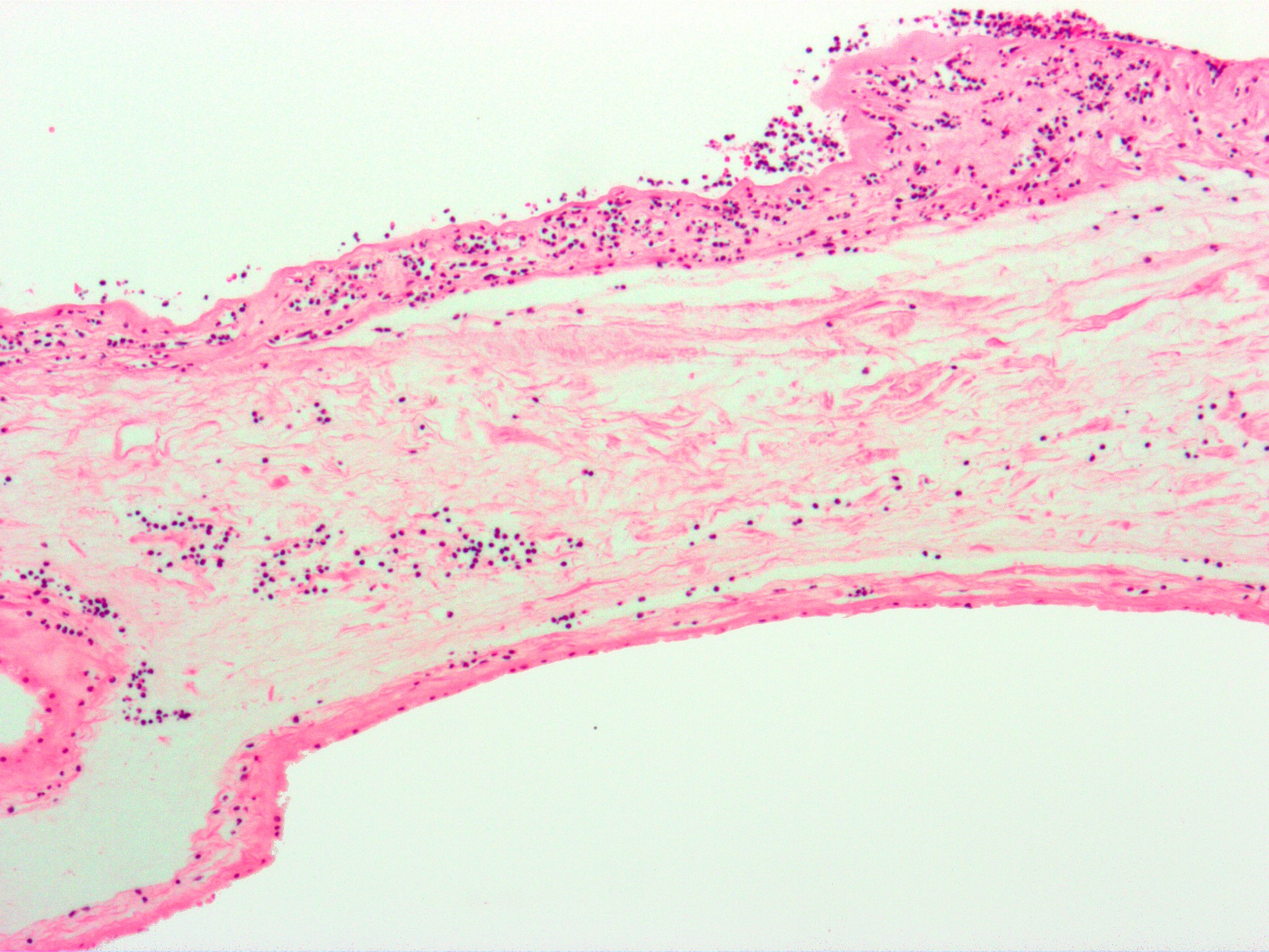
English: Micrograph of chorioamnionitis. H&E stain.
The amnion is seen at the very bottom of the image and composed of a simple cuboidal epithelium and a layer of eosinophilic (pink) connective tissue. It has a few scattered neutrophils - which makes the diagnosis of chorioamnionitis. Above the amnion is the chorion, which also has neutrophils. The fetus (baby) - not shown - would be below the image (and surrounded by amnionic fluid). The top of the image is the maternal aspect of the fetal membranes. It has significant inflammation. The pattern of inflammation seen in this image is typical of chorioamnionitis; most chorioamnionitis results from an ascending infection, i.e. the microorganisms ascend from the vagina/cervix. See also
|
||
| 来源 | 自己的作品 | ||
| 作者 | Nephron | ||
| 授权 (二次使用本文件) |
我,本作品著作权人,特此采用以下许可协议发表本作品: 本文件采用知识共享署名-相同方式共享 3.0 未本地化版本许可协议授权。
您可以选择您需要的许可协议。 |

文件历史
点击某个日期/时间查看对应时刻的文件。
| 日期/时间 | 缩略图 | 大小 | 用户 | 备注 | |
|---|---|---|---|---|---|
| 当前 | 2009年3月11日 (三) 09:27 | | 2,048 × 1,536(746 KB) | Nephron(留言 | 贡献) | {{Information |Description={{en|1=Micrograph of '''chorioamnionitis'''. H&E stain. The amnion is seen at the very bottom of the image and composed of a [[w:simple epithelium|simple cubo |
您不可以覆盖此文件。
文件用途
没有页面使用本文件。
全域文件用途
以下其他wiki使用此文件:
- es.wikipedia.org上的用途
- zh.wikipedia.org上的用途