File:Computed tomography of human brain - large.png
跳至導覽
跳至搜尋

預覽大小:800 × 570 像素。 其他解析度:320 × 228 像素 | 640 × 456 像素 | 1,024 × 730 像素 | 1,280 × 913 像素 | 2,560 × 1,826 像素 | 3,639 × 2,595 像素。
原始檔案 (3,639 × 2,595 像素,檔案大小:3.9 MB,MIME 類型:image/png)
說明
說明
添加單行說明來描述出檔案所代表的內容
Divya rai brain report
| 此檔案在創用CC CC0 1.0 通用公有領域貢獻宣告之下分發。 | |
| 在此宣告之下分發本作品者,已依據各國著作權法,在全世界放棄其對本作品所擁有的著作權及所有相關相似的法律權利,從而將本作品貢獻至公有領域。您可以複製、修改、分發和演示該作品,用於任何商業用途,所有這些都不需要請求授權。
|
|

| 描述 |
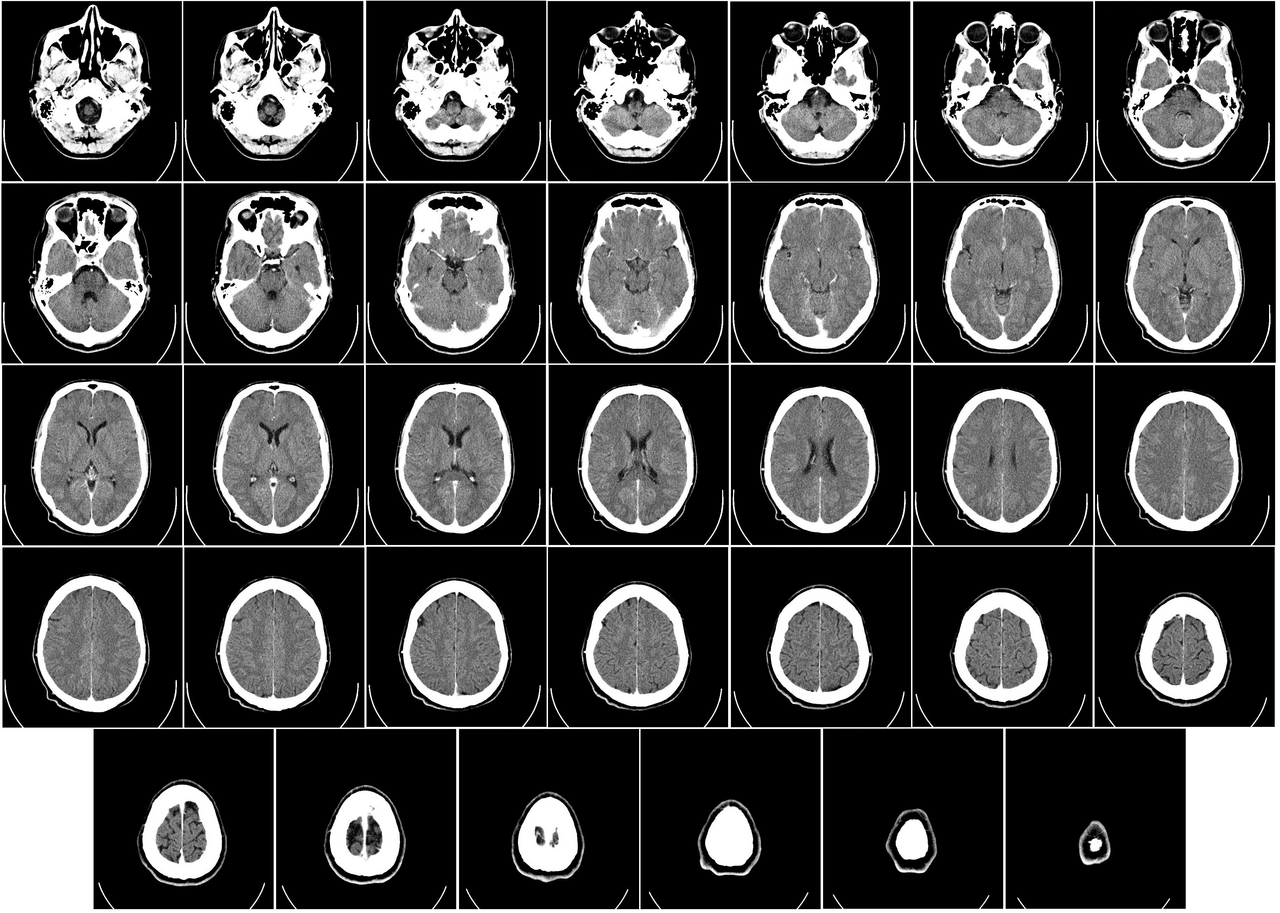
English: Computer tomography of human brain, from base of the skull to top. Taken with intravenous contrast medium.
It was taken Mars 23, 2007 on the uploader, after a 20 minute episode of homonymous hemianopsia with loss of the left visual field, but nothing strange was found. Three episodes of scotoma occurred in the following years, whereof the last one was scintillating (depiction). Otherwise, there were no further neurological symptoms.
Türkçe: Geçirdiği bir kaza neticesinde homonim hemianopsi vakası oluşan bir hastanın beyninin bilgisayarlı tomografisi. Tomografi neticesinde bir anomaliye rastlanmamıştır. |
| 日期 | Uploaded January 17, 2008 |
| 來源 | Radiology, Uppsala University Hospital. Uploaded by Mikael Häggström. |
| 作者 | Department of Radiology, Uppsala University Hospital. Uploaded by Mikael Häggström. |
| 授權許可 (重用此檔案) |
Compound images[編輯]
-

-
 Inverted


Scrollable stack[編輯]
For larger version, see Category:Computed tomography images of Mikael Häggström's brain. To move through the images, hover over the image and use scroll wheel, drag the mouse, or click the < or the > above each stack. This functionality should activate when the page is fully loaded, which may take some time.

.png)
.png)
.png)
.png)
.png)
.png)
.png)
.png)
.png)
.png)
.png)
.png)
.png)
.png)
.png)
.png)
.png)
.png)
.png)
.png)
.png)
.png)
.png)
.png)
.png)
.png)
.png)
.png)
.png)
.png)
.png)
.png)
.png)
.png)
Case with multiplanar reconstruction[編輯]
-
 Brain, case 1: Multiplanar, but no intravenous contrast.
Individual images[編輯]
Licencing[編輯]
| 此檔案在創用CC CC0 1.0 通用公有領域貢獻宣告之下分發。 | |
| 在此宣告之下分發本作品者,已依據各國著作權法,在全世界放棄其對本作品所擁有的著作權及所有相關相似的法律權利,從而將本作品貢獻至公有領域。您可以複製、修改、分發和演示該作品,用於任何商業用途,所有這些都不需要請求授權。
|
DICOM format[編輯]
檔案歷史
點選日期/時間以檢視該時間的檔案版本。
| 日期/時間 | 縮圖 | 尺寸 | 用戶 | 備註 | |
|---|---|---|---|---|---|
| 目前 | 2017年12月24日 (日) 01:11 | | 3,639 × 2,595(3.9 MB) | Shashi.(對話 | 貢獻) | Reverted to version as of 12:49, 1 February 2008 (UTC) |
| 2008年5月8日 (四) 10:59 |  | 3,639 × 2,595(3.17 MB) | CountingPine(對話 | 貢獻) | Optimise using PNGOUT | |
| 2008年2月1日 (五) 12:49 |  | 3,639 × 2,595(3.9 MB) | Mikael Häggström(對話 | 貢獻) | {{34 computer tomography images}} {{Individual images of CT of Mikael Häggström's brain}} | |
| 2008年1月31日 (四) 11:56 |  | 3,639 × 2,595(4.03 MB) | Mikael Häggström(對話 | 貢獻) | {{34 computer tomography images}} {{Individual images of CT of Mikael Häggström's brain}} |
無法覆蓋此檔案。
檔案用途
下列41個頁面有用到此檔案:
- User:Dronebogus/Favorites
- User:Mikael Häggström
- File:Anatomy image for main menu.png
- File:CT of brain of Mikael Häggström large.png (檔案重新導向)
- File:Computed tomography of human brain (1).png
- File:Computed tomography of human brain (10).png
- File:Computed tomography of human brain (11).png
- File:Computed tomography of human brain (12).png
- File:Computed tomography of human brain (13).png
- File:Computed tomography of human brain (14).png
- File:Computed tomography of human brain (15).png
- File:Computed tomography of human brain (16).png
- File:Computed tomography of human brain (17).png
- File:Computed tomography of human brain (18).png
- File:Computed tomography of human brain (19).png
- File:Computed tomography of human brain (2).png
- File:Computed tomography of human brain (20).png
- File:Computed tomography of human brain (21).png
- File:Computed tomography of human brain (22).png
- File:Computed tomography of human brain (23).png
- File:Computed tomography of human brain (24).png
- File:Computed tomography of human brain (25).png
- File:Computed tomography of human brain (26).png
- File:Computed tomography of human brain (27).png
- File:Computed tomography of human brain (28).png
- File:Computed tomography of human brain (29).png
- File:Computed tomography of human brain (3).png
- File:Computed tomography of human brain (30).png
- File:Computed tomography of human brain (31).png
- File:Computed tomography of human brain (32).png
- File:Computed tomography of human brain (33).png
- File:Computed tomography of human brain (34).png
- File:Computed tomography of human brain (4).png
- File:Computed tomography of human brain (5).png
- File:Computed tomography of human brain (6).png
- File:Computed tomography of human brain (7).png
- File:Computed tomography of human brain (8).png
- File:Computed tomography of human brain (9).png
- File:Computed tomography of human brain - large, inverted.png
- File:Computed tomography of human brain - large.png
- Template:34 computer tomography images
全域檔案使用狀況
以下其他 wiki 使用了這個檔案:
- bn.wikipedia.org 的使用狀況
- bo.wikipedia.org 的使用狀況
- ca.wikipedia.org 的使用狀況
- en.wikipedia.org 的使用狀況
- CT scan
- Portal:Medicine
- Portal:Medicine/Selected picture
- Portal:Medicine/Selected picture archive
- Wikipedia:WikiProject Neuroscience
- Wikipedia:Featured pictures/Sciences/Biology
- User:Mikael Häggström
- User talk:Mikael Häggström/Archive 1
- Wikipedia:Featured pictures thumbs/10
- Wikipedia:Featured picture candidates/CT of brain of Mikael Häggström.png
- Wikipedia:Featured picture candidates/February-2008
- Wikipedia:Wikipedia Signpost/2008-02-11/Features and admins
- Portal:Medicine/Selected picture/9, 2008
- Portal:Medicine/Selected picture/9
- Wikipedia:Picture of the day/July 2008
- Template:POTD/2008-07-11
- Wikipedia:Wikipedia Signpost/2008-02-11/SPV
- User:Mikael Häggström/Gallery
- Wikipedia:WikiProject Medicine/Recognized content
- Computed tomography of the head
- Wikipedia:Wikipedia Signpost/2013-10-02/Op-ed
- Wikipedia:Wikipedia Signpost/Single/2013-10-02
- User:Wouterstomp/test
- User:Fitness queen04/sandbox
- Wikipedia:WikiProject Anatomy/Resources
- Wikipedia:WikiProject Anatomy/Recognized content
- Wikipedia talk:WikiProject Anatomy/Archive 9
- Reconstruction from projections
- User:VGrigas (WMF)/Quality Media
- User:Flyer22 Frozen/Human brain
- Portal:Medicine/Recognized content
- User talk:Rhododendrites/Reconsidering FPC on the English Wikipedia
- es.wikipedia.org 的使用狀況
- fi.wikipedia.org 的使用狀況
- he.wikipedia.org 的使用狀況
- hy.wikipedia.org 的使用狀況
- hyw.wikipedia.org 的使用狀況
- id.wikipedia.org 的使用狀況
- is.wikipedia.org 的使用狀況
- ja.wikipedia.org 的使用狀況
檢視此檔案的更多全域使用狀況。